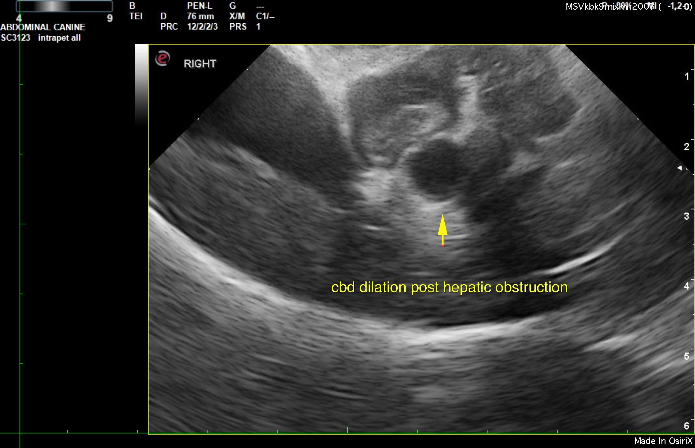
The right limb of the pancreas extended around the common bile duct causing dilation of the common bile duct at approximately 0.8 cm. Post hepatic obstruction is present.
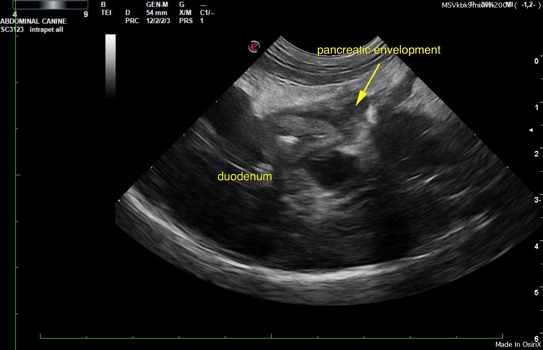
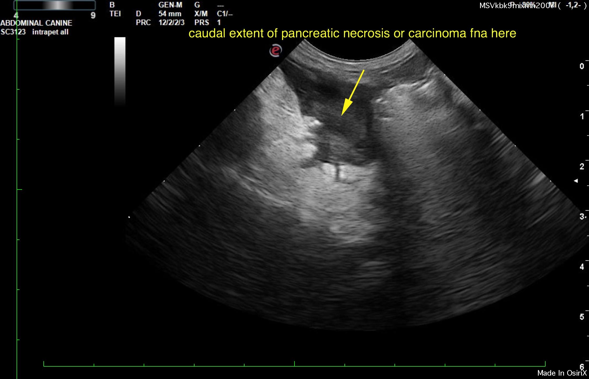
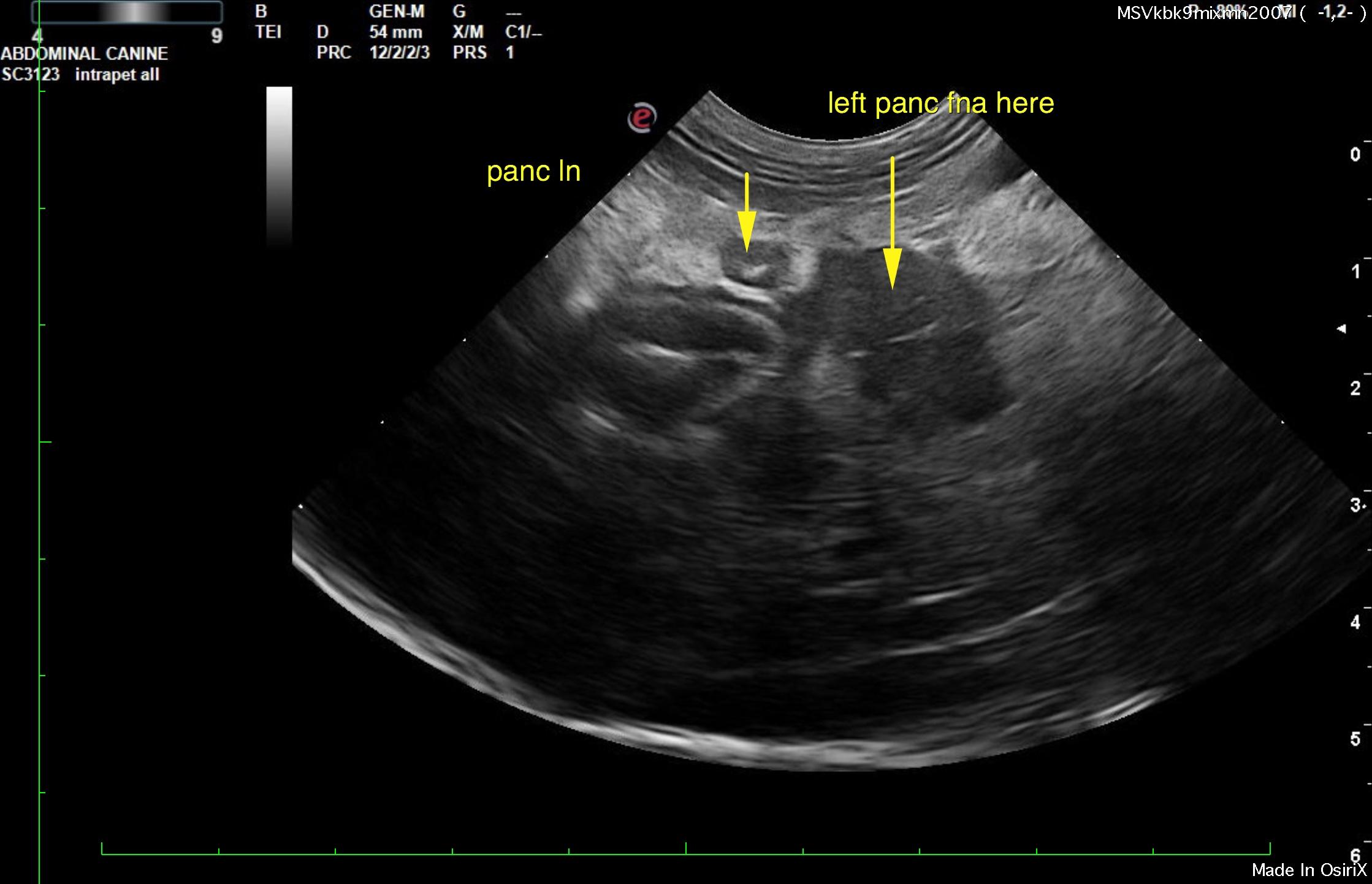
The pancreas revealed mixed, hypoechoic and hyperechoic changes with regional lymphadenopathy. Ultrasound-guided FNA is strongly recommended to rule out pancreatic carcinoma versus necrosis and pancreatitis. See image below with targets to sample. The pancreatic pathology extended caudally towards the colon with regional adhesions. This is consistent with pancreatic necrosis. Culture of the fluid and tissue obtained from this region is recommended. The right limb of the pancreas extended around the common bile duct causing dilation of the common bile duct at approximately 0.8 cm. The regional lymph node was enlarged and measured 0.5 cm.
Regional free fluid was noted.