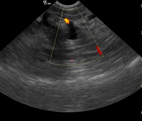
A 14-year-old MN DSH cat was presented for anorexia and lack of defecation. On physical examination, the eyes were sunken and the abdomen was soft and doughy. CBC showed leukocytosis, neutrophilia, lymphopenia, monocytosis, eosinopenia, and mild thrombocytosis. The only pertinent finding on blood chemistry was hyperglycemia. T-4 was within normal range. The patient was treated with subcutaneous fluids and force-fed A/D. Two days later the patient was hospitalized and treated with I.V. fluids, antibiotics, and supportive care. SPEC FPL result was high.
A 14-year-old MN DSH cat was presented for anorexia and lack of defecation. On physical examination, the eyes were sunken and the abdomen was soft and doughy. CBC showed leukocytosis, neutrophilia, lymphopenia, monocytosis, eosinopenia, and mild thrombocytosis. The only pertinent finding on blood chemistry was hyperglycemia. T-4 was within normal range. The patient was treated with subcutaneous fluids and force-fed A/D. Two days later the patient was hospitalized and treated with I.V. fluids, antibiotics, and supportive care. SPEC FPL result was high.