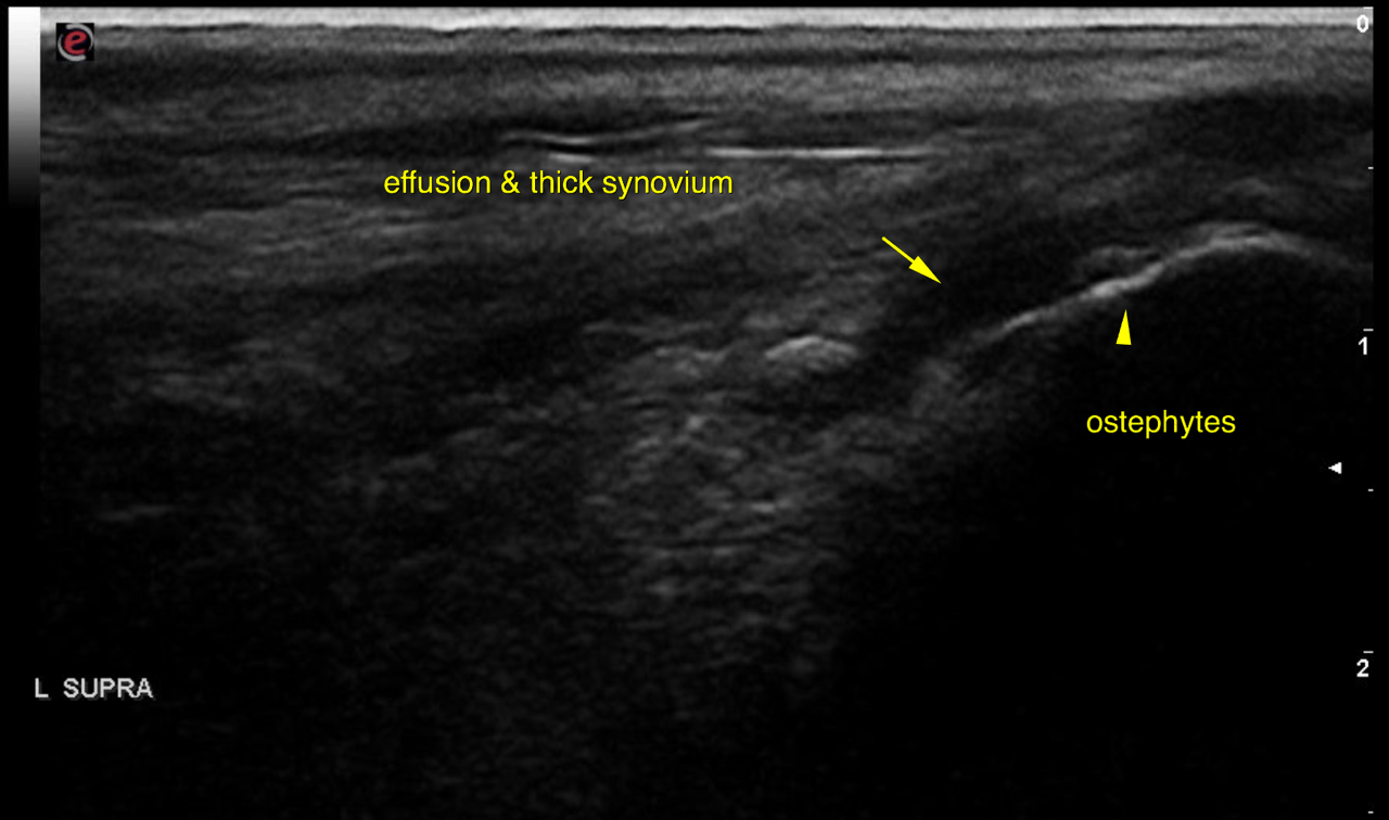
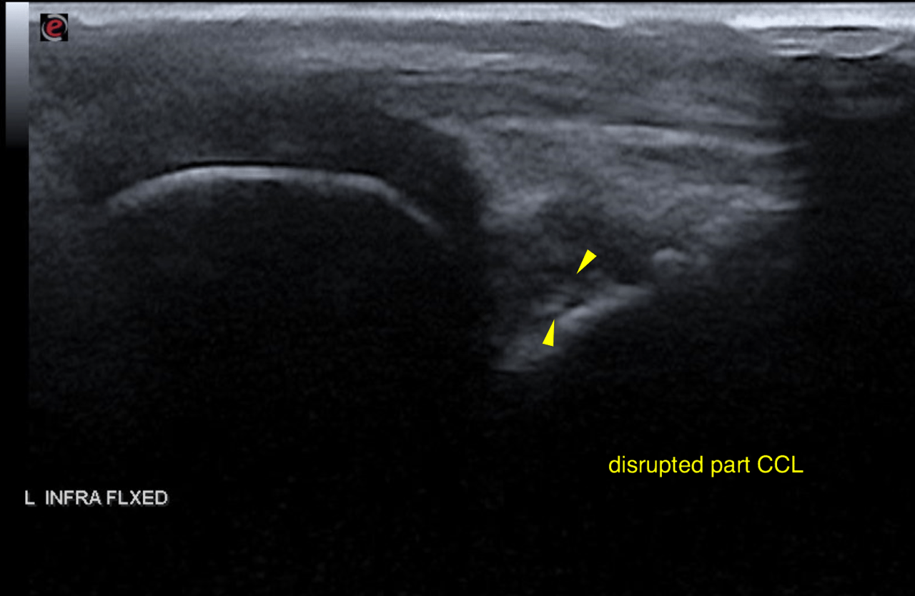
History of right hind lameness at the beginning of January 2016. Left and right knee rads and pelvis unremarkable at the time. No cranial drawer sign noted on either knee. Pet was placed on a trial of Metacam but owner still seeing lameness. Pet re-examined today and lameness noted on the L hind leg. Crepitus noted in left knee and subtle cranial drawer present. Ultrasound of left and right knees performed.
History of right hind lameness at the beginning of January 2016. Left and right knee rads and pelvis unremarkable at the time. No cranial drawer sign noted on either knee. Pet was placed on a trial of Metacam but owner still seeing lameness. Pet re-examined today and lameness noted on the L hind leg. Crepitus noted in left knee and subtle cranial drawer present. Ultrasound of left and right knees performed.