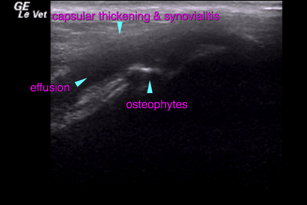
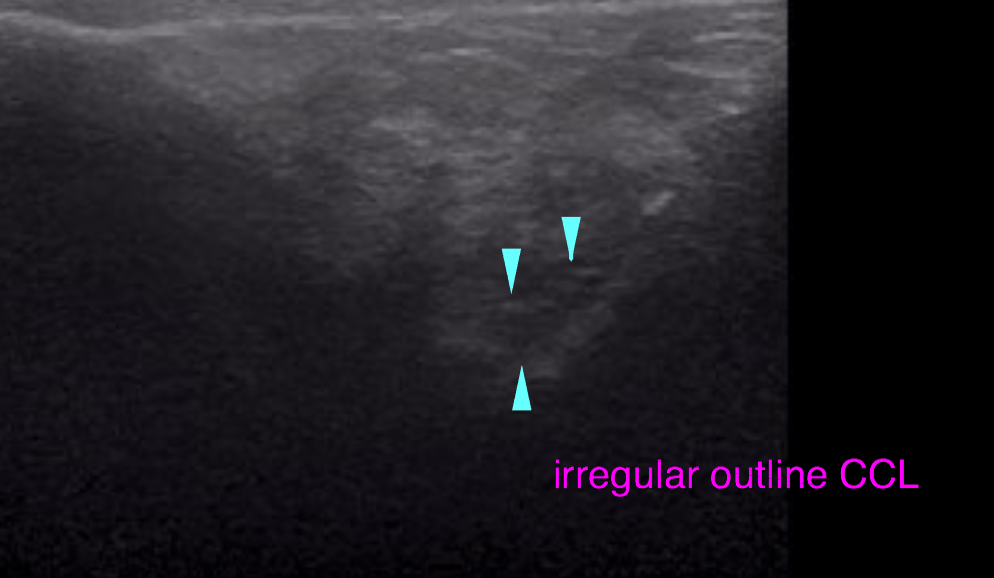
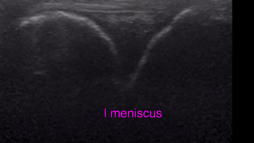
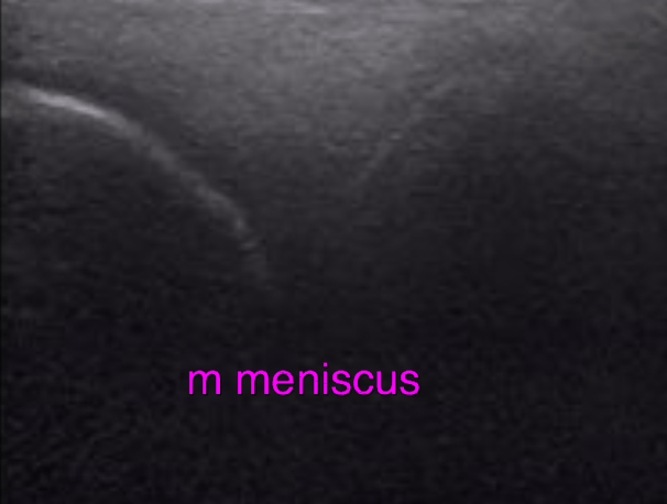
Ultrasound of the left stifle –
Moderate thickening of the joint capsule with synovialitis is noted. A moderate amount of osteophytes is seen at the periarticular margins. The femoropatellar joint cartilage presents mild irregularity at the most proximal extent of the femoral trochlea. The infrapatellar fat pad presents an irregular echo pattern. Most of the cranial cruciate ligament (CCL) fibres are continuous but with increased echogenicity and irregular delineation. The continuous fibres appear to be elongated. The medial and lateral meniscus are ultrasonographically within normal limits.