ORTHO – Partial Cranial Cruciate Ligament (CCL) Rupture and Chronic Degenerative Joint Disease in a 6 year old Brittany Spaniel dog

Case Study

ORTHO – Partial Cranial Cruciate Ligament (CCL) Rupture and Chronic Degenerative Joint Disease in a 6 year old Brittany Spaniel dog

History of left hind lameness. Physical exam showed grade 2 cranial drawer.

History of left hind lameness. Physical exam showed grade 2 cranial drawer.

Image Interpretation

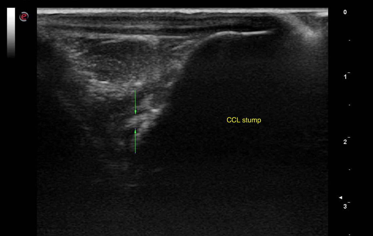
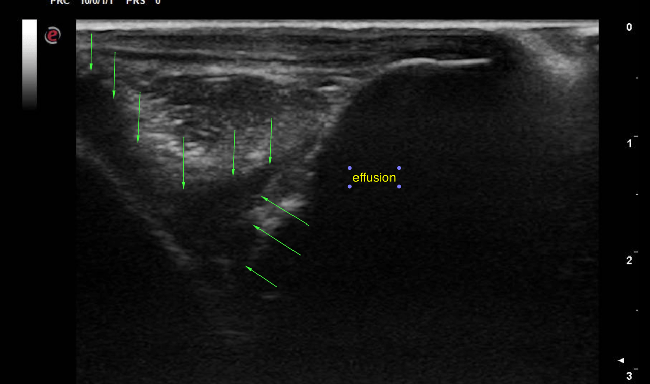
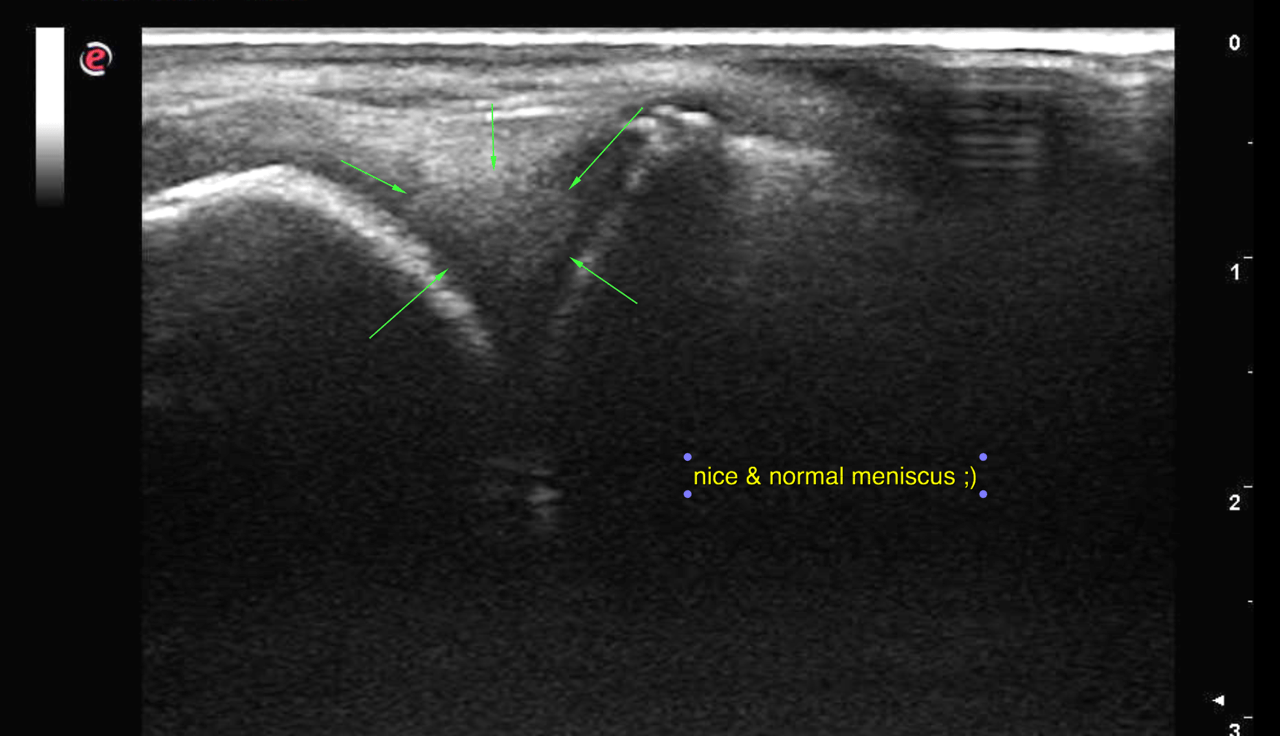
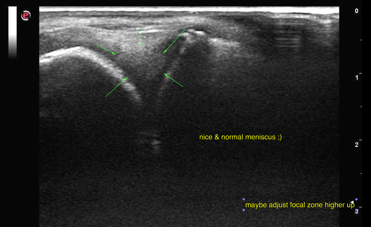
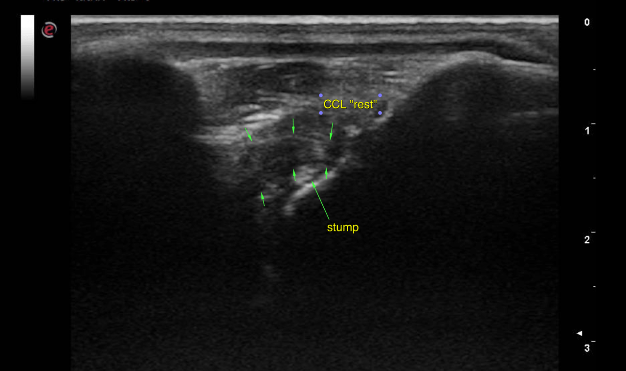
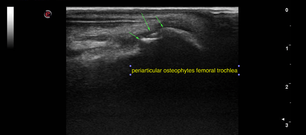
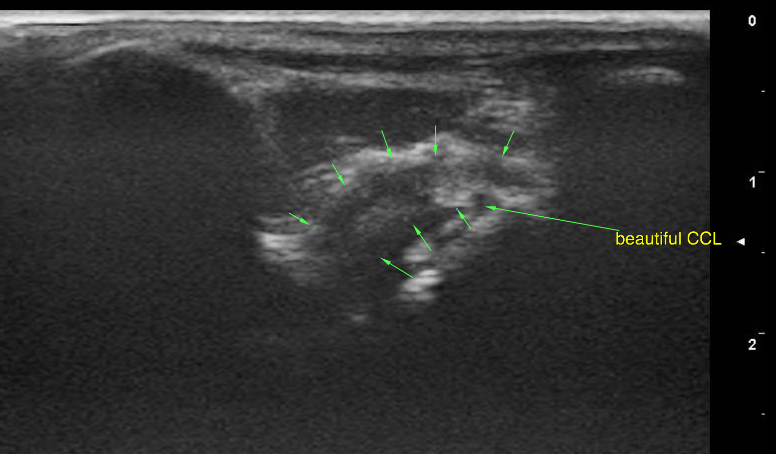
Ultrasound of the left stifle – The left stifle joint presented moderate anechoic effusion, moderate capsular thickening and moderate synovial proliferation. There were mild to moderate osteophyte formations at the periarticular margins. The femoral condylar cartilage surface appeared to be mildly roughened. The cranial cruciate ligament (CCL) presented partial loss of integrity with irregular margination, uneven thickness and periligamentous effusion. There was a small hyperechoic fibre stump visible next to the distal insertion of the CCL at the tibial intercondylar eminence. The caudal cruciate ligament was not seen. The medial and lateral meniscus did not present ultrasonographic abnormalities.
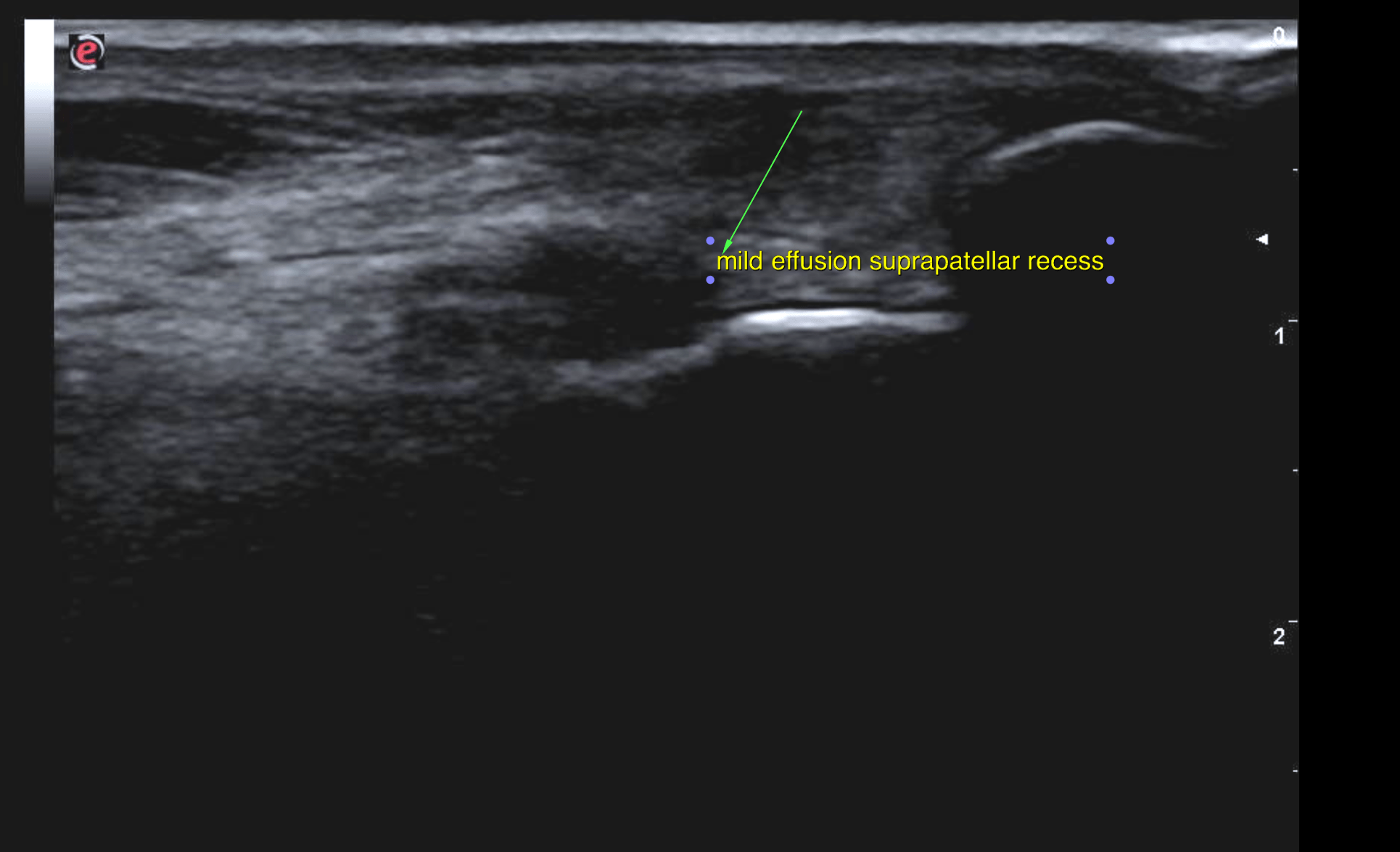
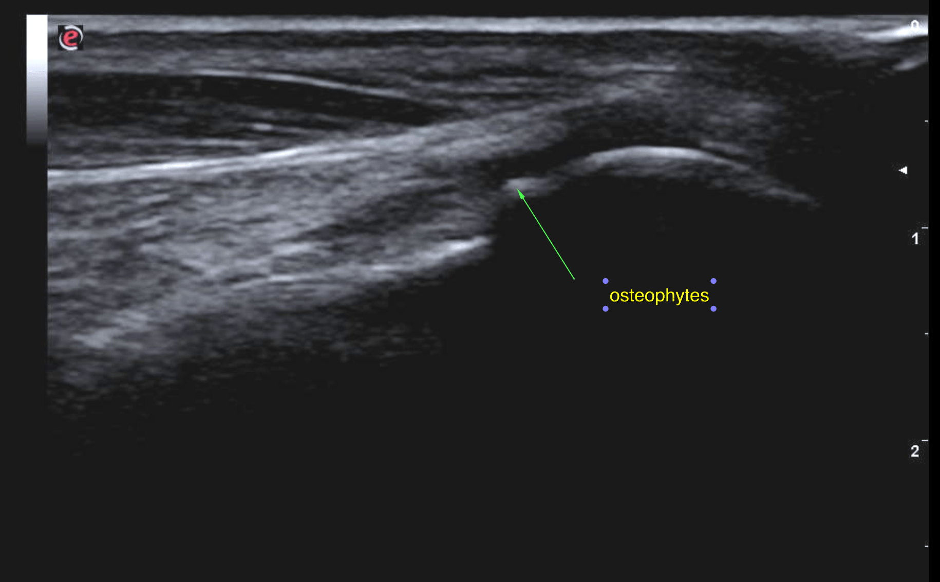
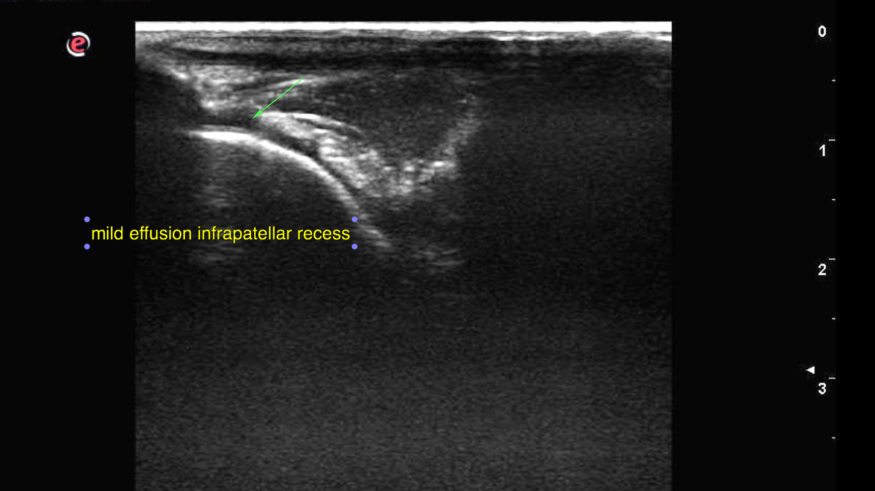
Ultrasound of the right stifle – The right stifle joint presented mild anechoic effusion within the supra- and infrapatellar recesses, mild capsular thickening and mild synovial proliferation. There were mild osteophyte formations at the periarticular margins. The cranial cruciate ligament (CCL) was continuous and even in thickness but presented minor echotextural and echogenicity changes. There was scant periligamentous effusion. The caudal cruciate ligament was not seen. The medial and lateral meniscus did not present ultrasonographic abnormalities.

DX

The ultrasonographic findings are compatible with a left-sided partial CCL rupture and chronic degenerative joint disease with moderate osteoarthrosis and synovialitis. In the right stifle there is mild chronic degenerative joint disease with mild osteoarthrosis and synovitis. The findings of the right cranial cruciate ligament are suggestive of edema and degeneration.

Patient Information

Patient Name : Bella McClean, Bayshore AH. Yay Dr. Hunt!
Species : Canine
Type of Imaging : Ultrasound
Status : Complete

Clinical Signs

  • Decreased Drinking

Images

bildschirmfoto_2015-08-11_um_11bildschirmfoto_2015-08-11_um_11bildschirmfoto_2015-08-11_um_11bildschirmfoto_2015-08-11_um_11bildschirmfoto_2015-08-11_um_11bildschirmfoto_2015-08-11_um_11bildschirmfoto_2015-08-13_um_20bildschirmfoto_2015-08-13_um_20bildschirmfoto_2015-08-13_um_20bildschirmfoto_2015-08-13_um_20bildschirmfoto_2015-08-13_um_20

Clinical Signs

  • Decreased Drinking
Skip to content