History of left hind lameness of 3 weeks duration. Toe touching. Physical exam: slightly swollen left stifle area.
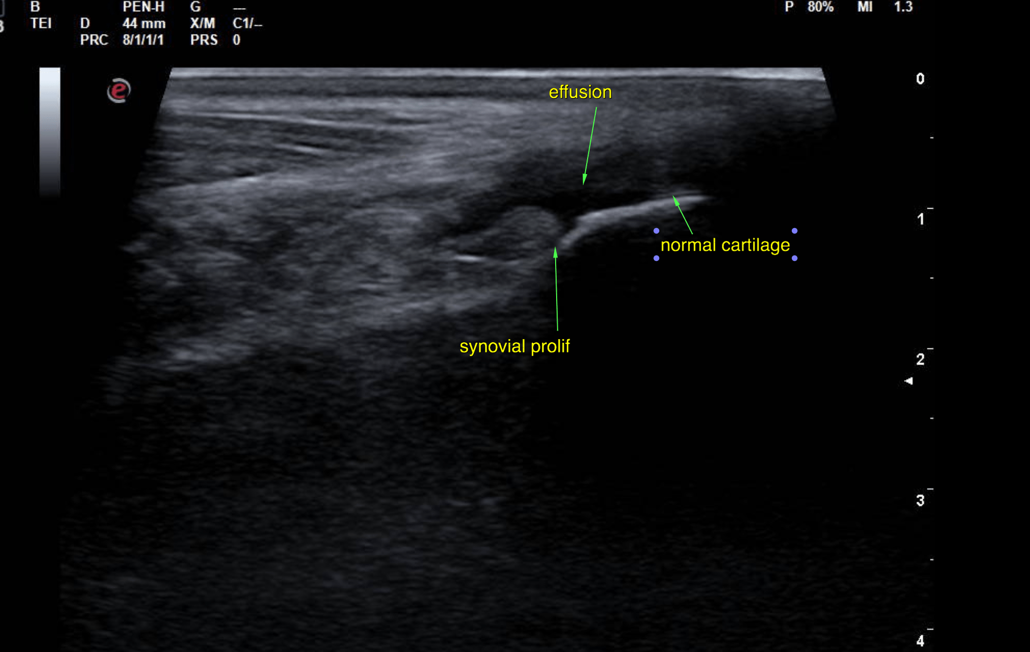
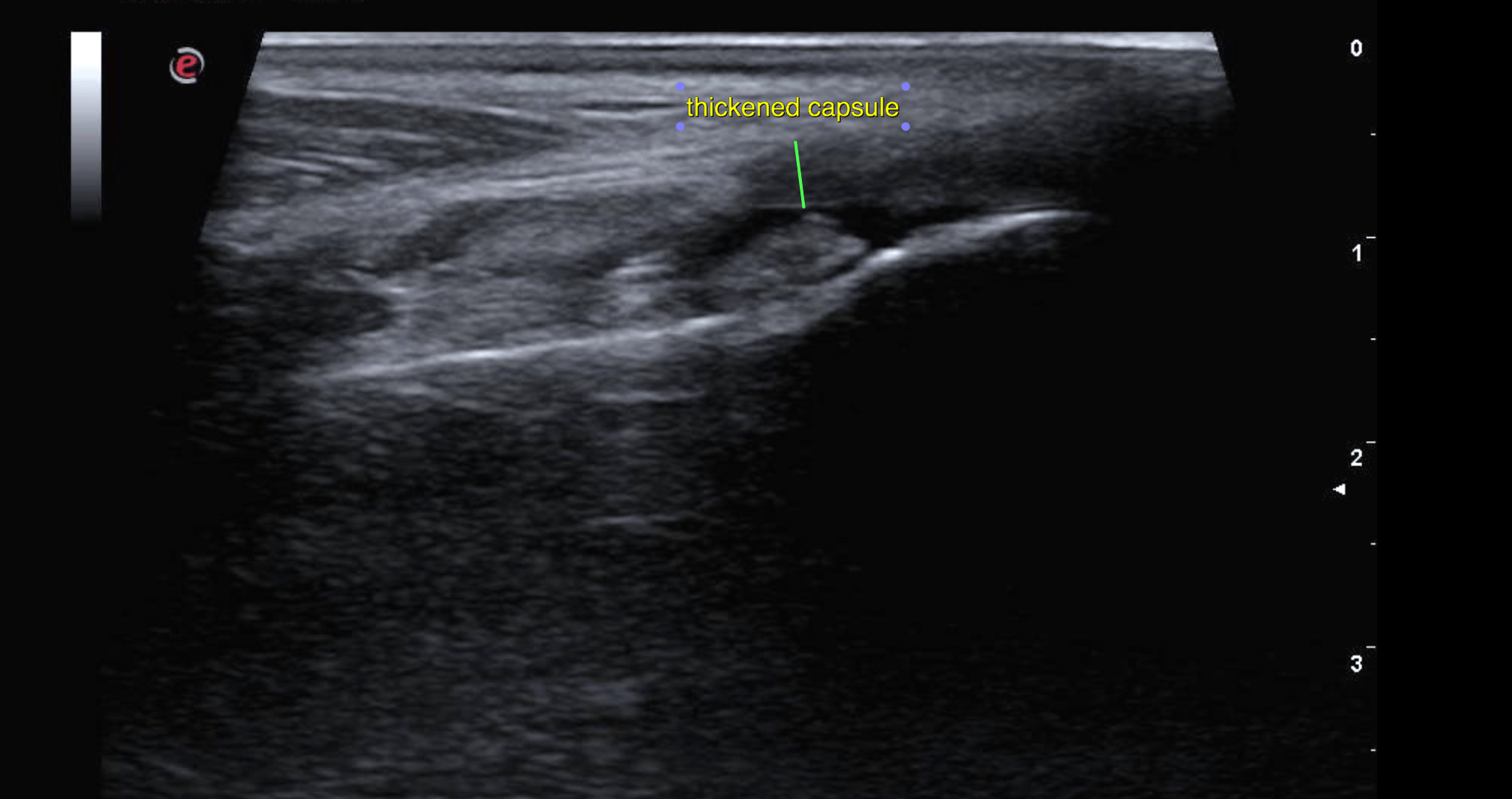
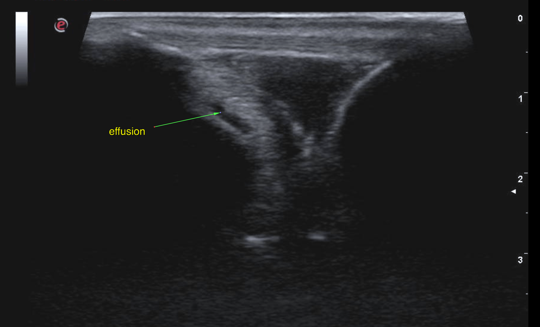
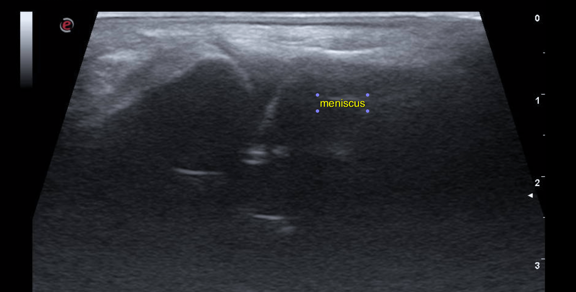
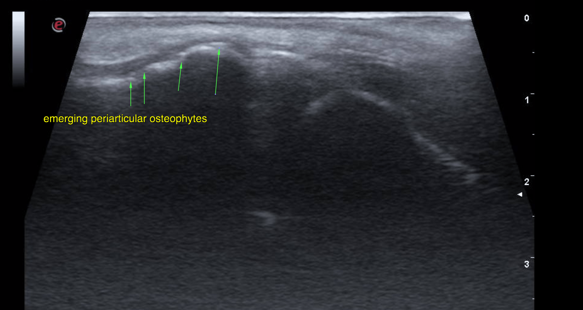
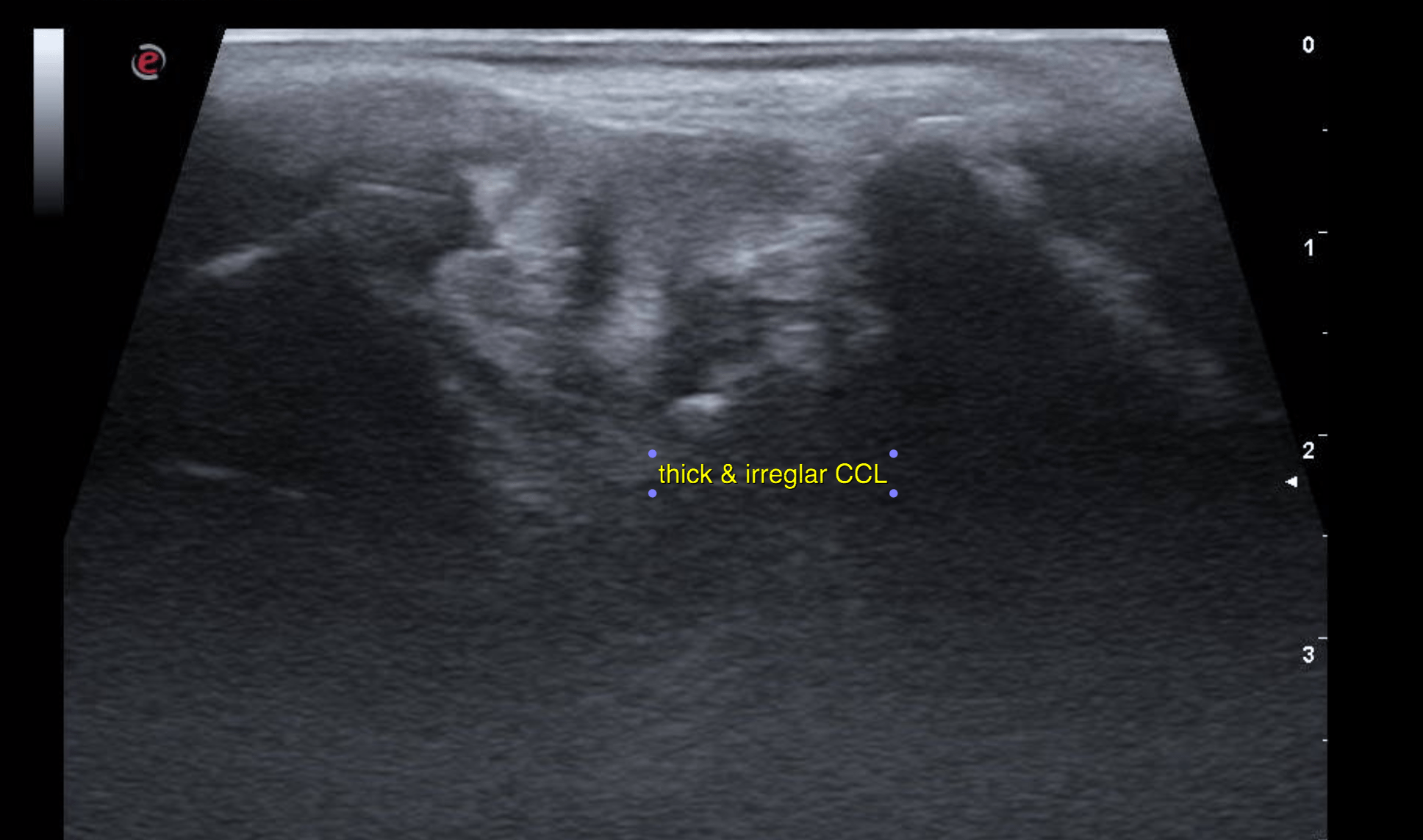
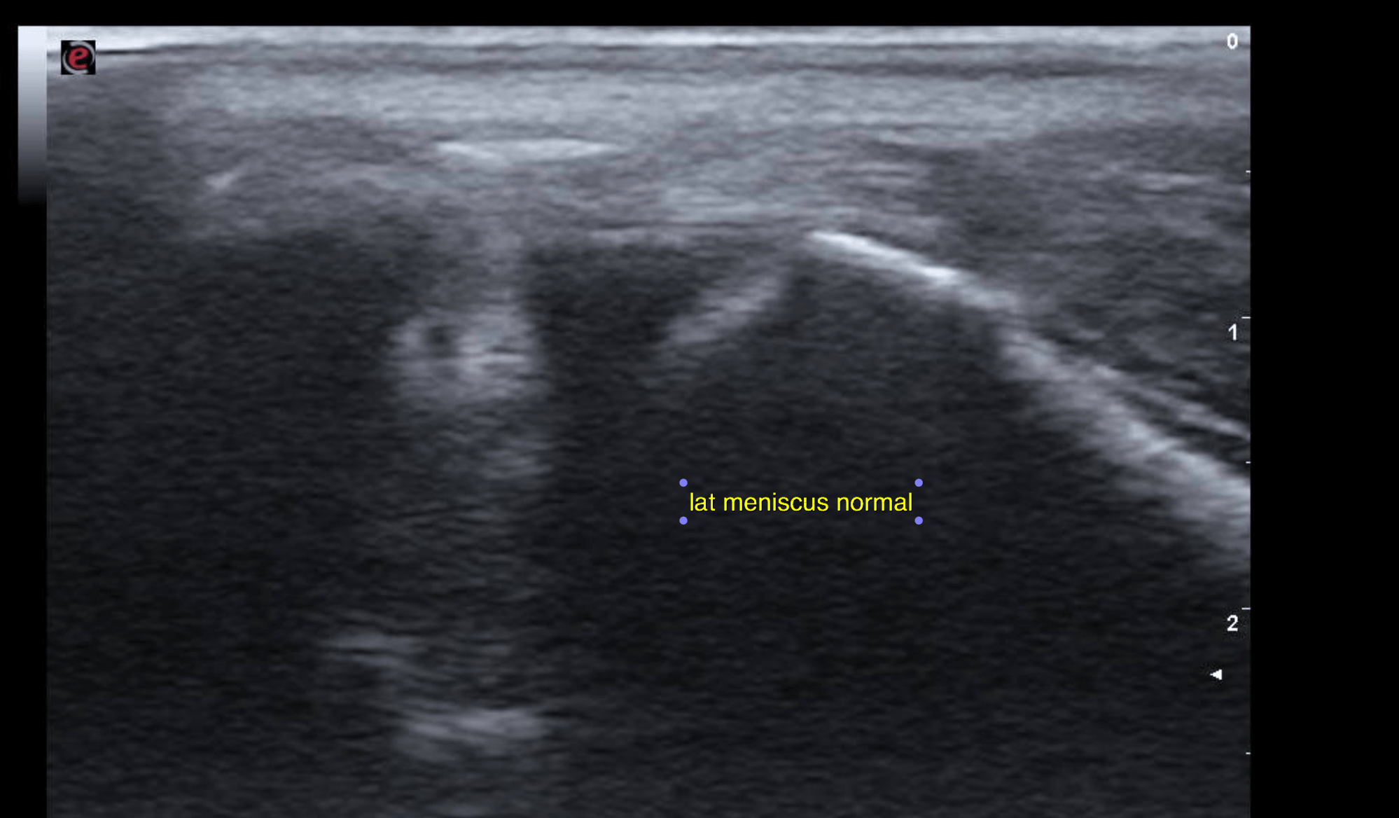
Ultrasound showed the left stifle joint presented moderate anechoic effusion, significant capsular thickening and significant synovial proliferation. There were emerging osteophyte formations at the periarticular margins. The femoral condylar cartilage surface was normal. The cranial cruciate ligament (CCL) presented partial loss of integrity with irregular margination, unevenly increased thickness and scant periligamentous effusion. The caudal cruciate ligament was not seen. The medial and lateral meniscus did not present ultrasonographic abnormalities.
A partial and relatively recent tear may explain the severity of the lameness.
Synovial aspiration is recommended to rule out differential diagnoses such as non-erosive arthritis since the signs of cartilage and bone degeneration are very mild in relation to the significant degree of synovial proliferation and inflammation.