ORTHO – normal shoulder musculature and tendons in a 2 year old FS Shetland Sheepdog with lameness

Case Study

ORTHO – normal shoulder musculature and tendons in a 2 year old FS Shetland Sheepdog with lameness

This 2 year old FS Shetland Sheepdog has a history of right ulnar osteotomy and corrective radial dome osteotomy 6 months previously. 
Previous conservative management of a right proximal diaphsyseal radius/ulna fracture sustained one year ago. Malunion resulting in caudal bowing of the bones.

This 2 year old FS Shetland Sheepdog has a history of right ulnar osteotomy and corrective radial dome osteotomy 6 months previously. 
Previous conservative management of a right proximal diaphsyseal radius/ulna fracture sustained one year ago. Malunion resulting in caudal bowing of the bones.

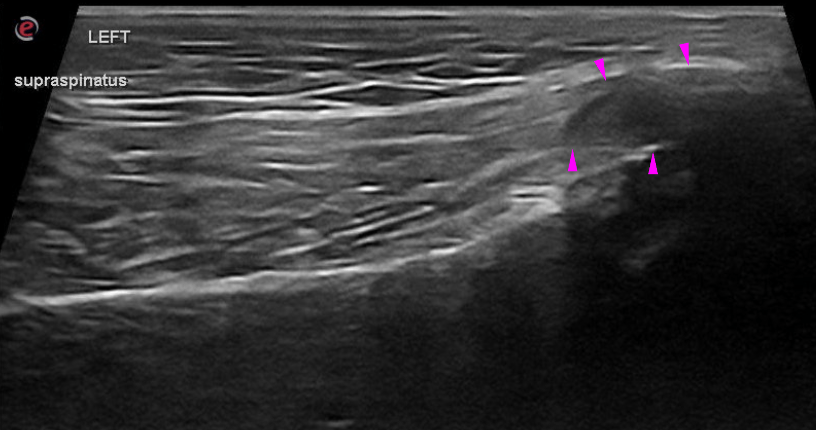
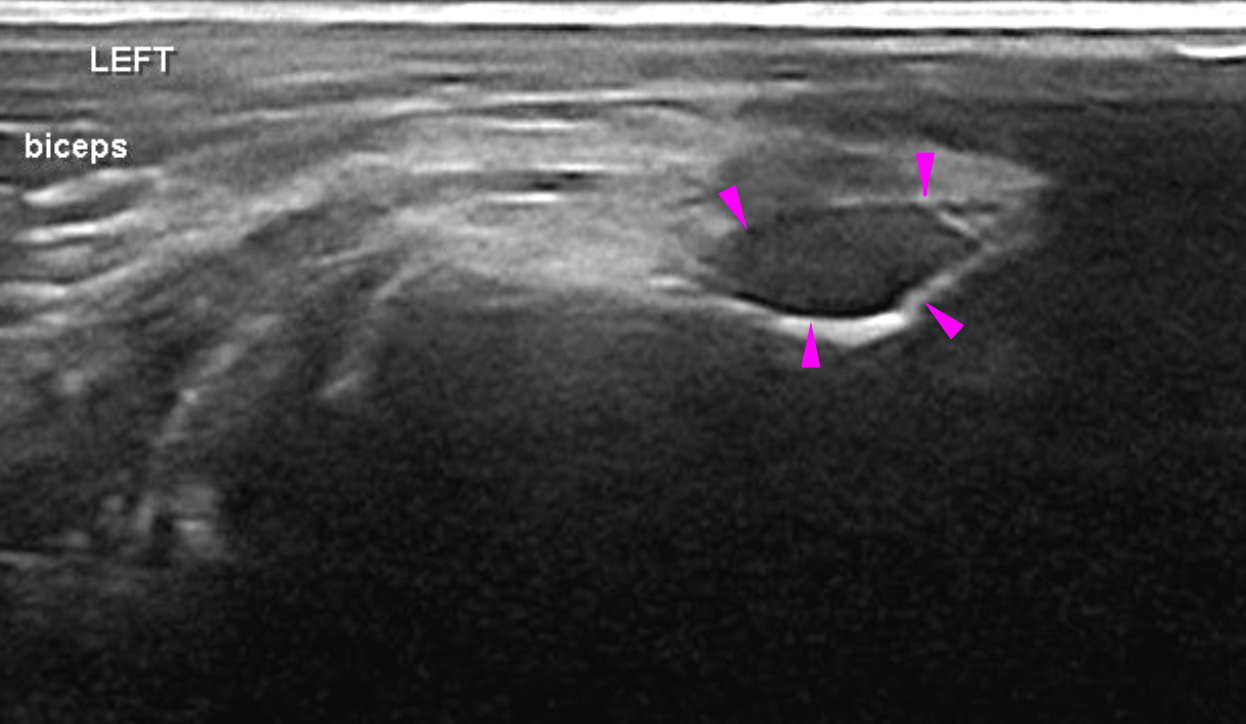
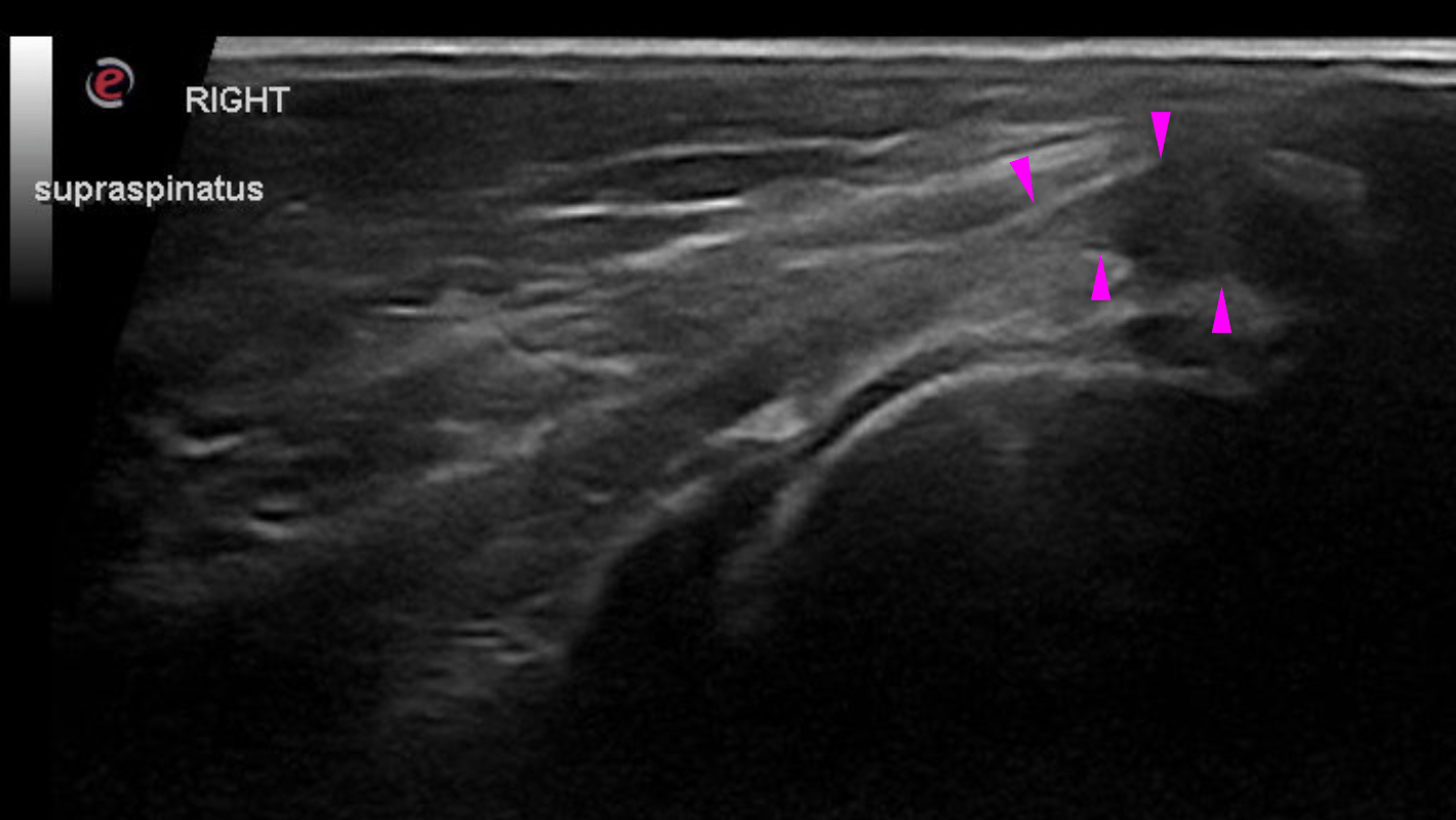
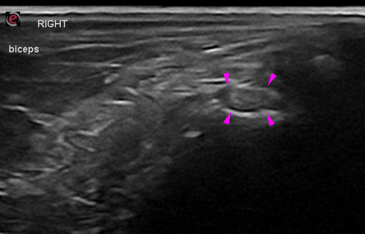
Physical exam: Gait/Lameness: Grade I-II/V lame in the right thoracic limb at a walk
; Posture: Stands square to slightly off weights the right thoracic limb while standing (buckles forward on the carpus)
; Muscle mass: Mild to moderate muscle atrophy over the right shoulder; 
Muscle palpation: Mild bicipital tendon spasm
; Joint palpation: All joints of the thoracic limbs have normal range of motion. Subjective increased abduction angle of both shoulders, more pronounced on the right. 
Ante brachium palpation: Overall plate quiet. Caudal aspect of ulna can be palpated. Both regions have mild sensitivity on deep palpation.       

Image Interpretation

Rads of right and left shoulder – 

The biceps tendon is well delineated, regular in diameter and presents with uniform echogenicity and echotexture. There is no tendon sheath effusion, no joint effusion and no thickening of the synovial lining noted. The intertubercular groove surface is even and smooth.

The distal insertion tendon of the supraspinatus presents with subjectively normal volume (equal volume between left and right). The greater humeral tubercle is even and smooth.

The infraspinatus muscle presents the expected echogenicity and fibre pattern. The thin distal insertion tendon is even in diameter, well-delineated and uniformy hyperechoic. Nu bursa enlargement is seen.

DX

The findings of the right and left shoulder musculature and tendons are within normal limits

Outcome

A pathology in the shoulder musculature can be ruled out as underlying cause of the current clinical presentation.

Patient Information

Patient Name : Olivia Winters
Gender : Female, Spayed
Species : Canine
Type of Imaging : Ultrasound
Status : Complete
Liz Wuz Here : Yes
Code : 18_00010

Clinical Signs

  • Lameness

Exam Finding

  • Muscle Wasting

Images

bildschirmfoto_2016-06-02_um_09bildschirmfoto_2016-06-02_um_09bildschirmfoto_2016-06-02_um_09bildschirmfoto_2016-06-02_um_09

Clinical Signs

  • Lameness