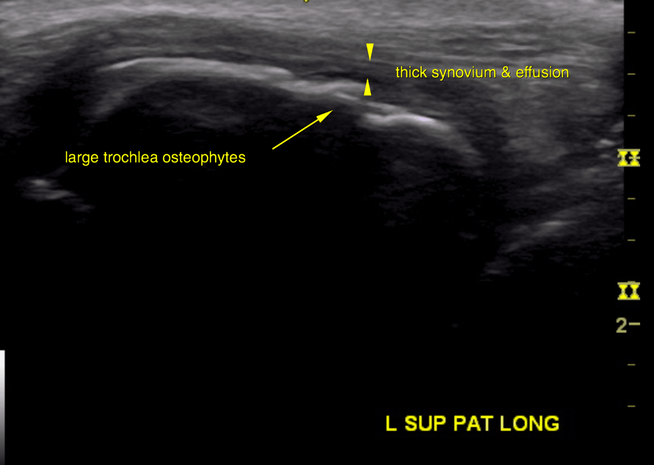
This 6 year old FS Nova Scotia Duck Tolling Retriever dog presented with injury to the left stifle. Positive cranial drawer, positive cranial tibial thrust (CTT), painful left stifle, and medial buttress.
CBC/Chem WNL
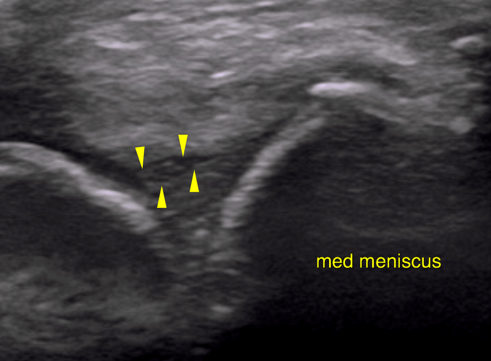
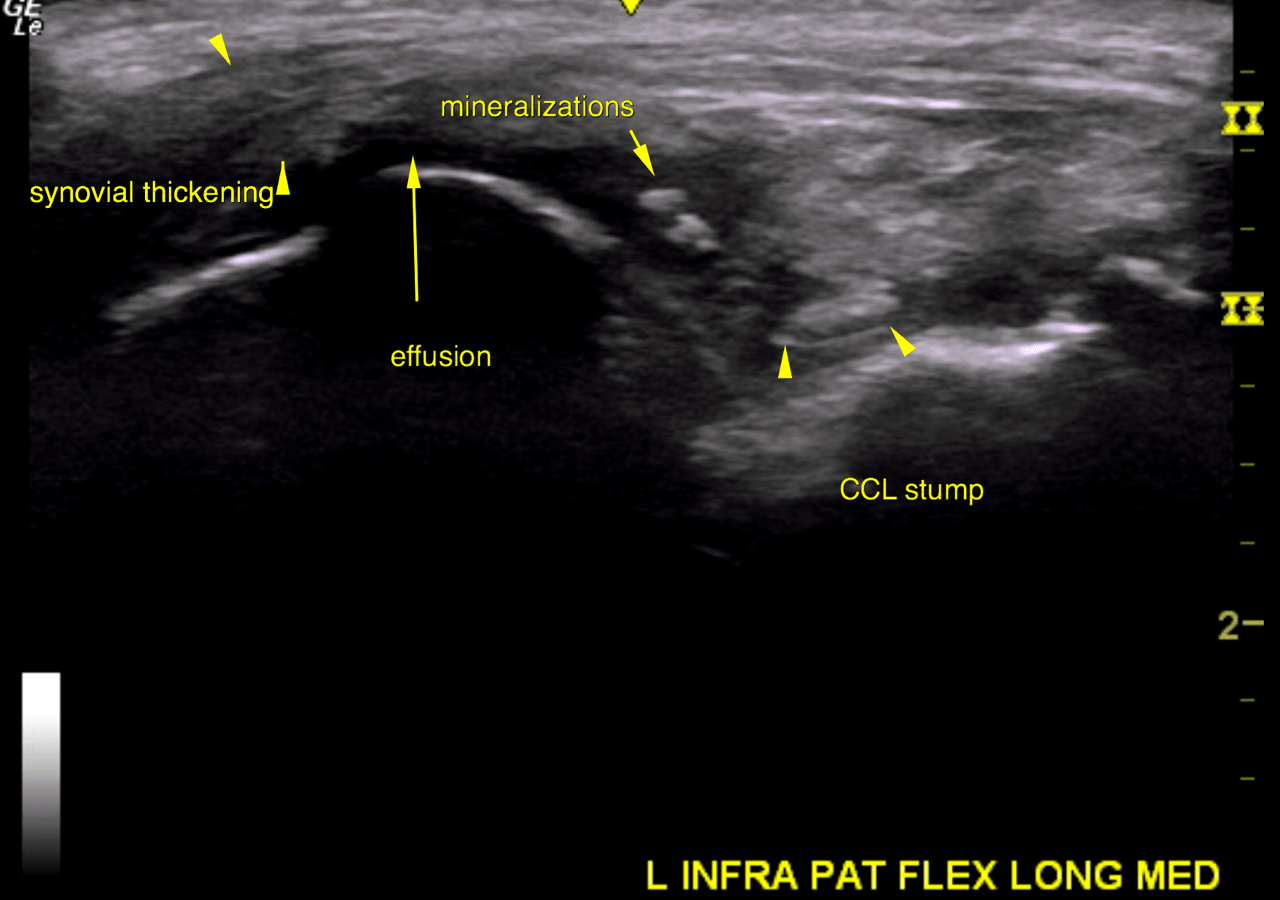
This 6 year old FS Nova Scotia Duck Tolling Retriever dog presented with injury to the left stifle. Positive cranial drawer, positive cranial tibial thrust (CTT), painful left stifle, and medial buttress.
CBC/Chem WNL