ORTHO – Degenerative joint disease (DJD) with edema of the cranial cruciate ligament (CCL) in a 4 year old MN Labrador Retriever

Case Study

ORTHO – Degenerative joint disease (DJD) with edema of the cranial cruciate ligament (CCL) in a 4 year old MN Labrador Retriever

This 4 year old MN Labrador Retriever dog has left hind lameness, suspect partial CCL tear

This 4 year old MN Labrador Retriever dog has left hind lameness, suspect partial CCL tear

Image Interpretation

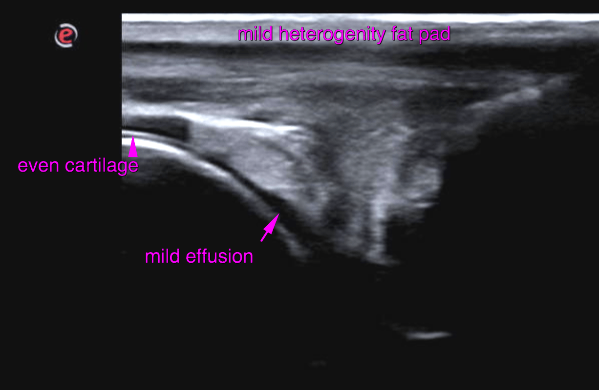
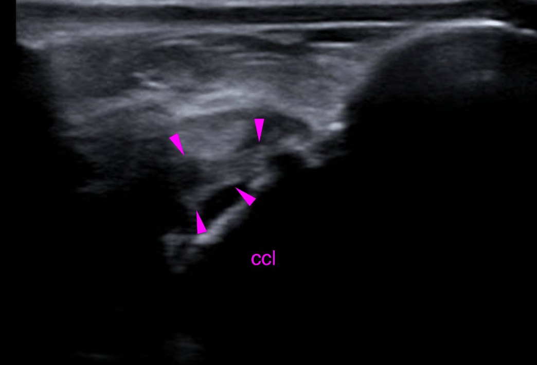
Ultrasound of the left stifle – The infrapatellar recess of the left stifle joint reveals mild synovial thickening as well as mild anechoic effusion. The cranial cruciate ligament (CCL) is well delineated, the regular fibre pattern is recongnized. The only abnormalities noted are a mildly uneven diameter and few echogenic foci. Mild periligamentous effusion and mild regional synovial membrane thickening are seen. The infrapatellar fat body presents mild heterogeneity as found commonly in degenerative joint disease (DJD).
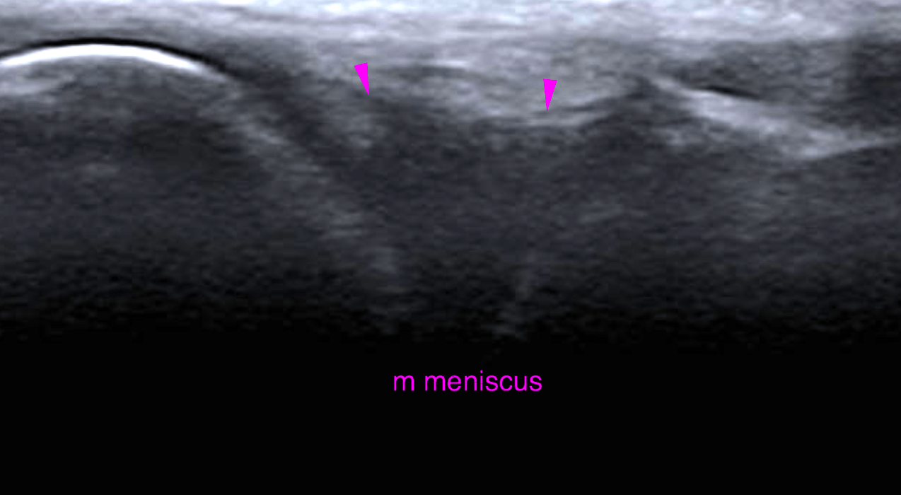
 The medial meniscus is in situ with regular outline and echogenicity

DX

DJD and edema of the CCL

Outcome

The ultrasonographic findings of the left stifle joint do not confirm the presence of partial or complete tearing of the cranial crucitae ligament at this point. The changes are subtle and support degeneration & edema of the cranial cruciate ligament which is a common prestage of cranial cruciate ligament pathology. However, biomechanical failure of the ligament is not very likely at present. Consider conservative management with clinical and ultrasonographic reevaluation in 4-8 weeks.

Patient Information

Patient Name : Otis Reevs/Bayshore
Gender : Male, Neutered
Species : Canine
Type of Imaging : Ultrasound
Status : Complete
Liz Wuz Here : Yes
Code : 18_00015

Clinical Signs

  • Lameness

Images

bildschirmfoto_2016-06-11_um_12bildschirmfoto_2016-06-11_um_12bildschirmfoto_2016-06-11_um_13bildschirmfoto_2016-06-11_um_13

Clinical Signs

  • Lameness
Skip to content