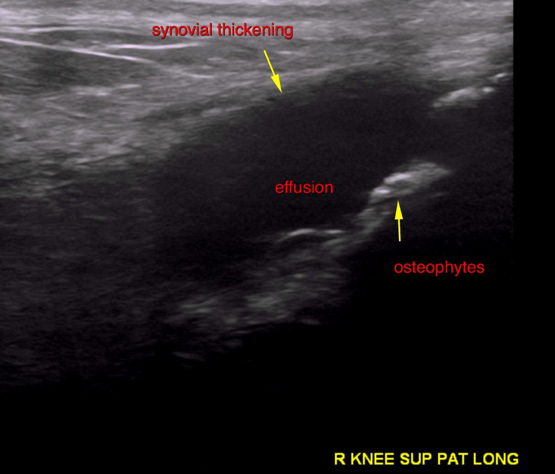
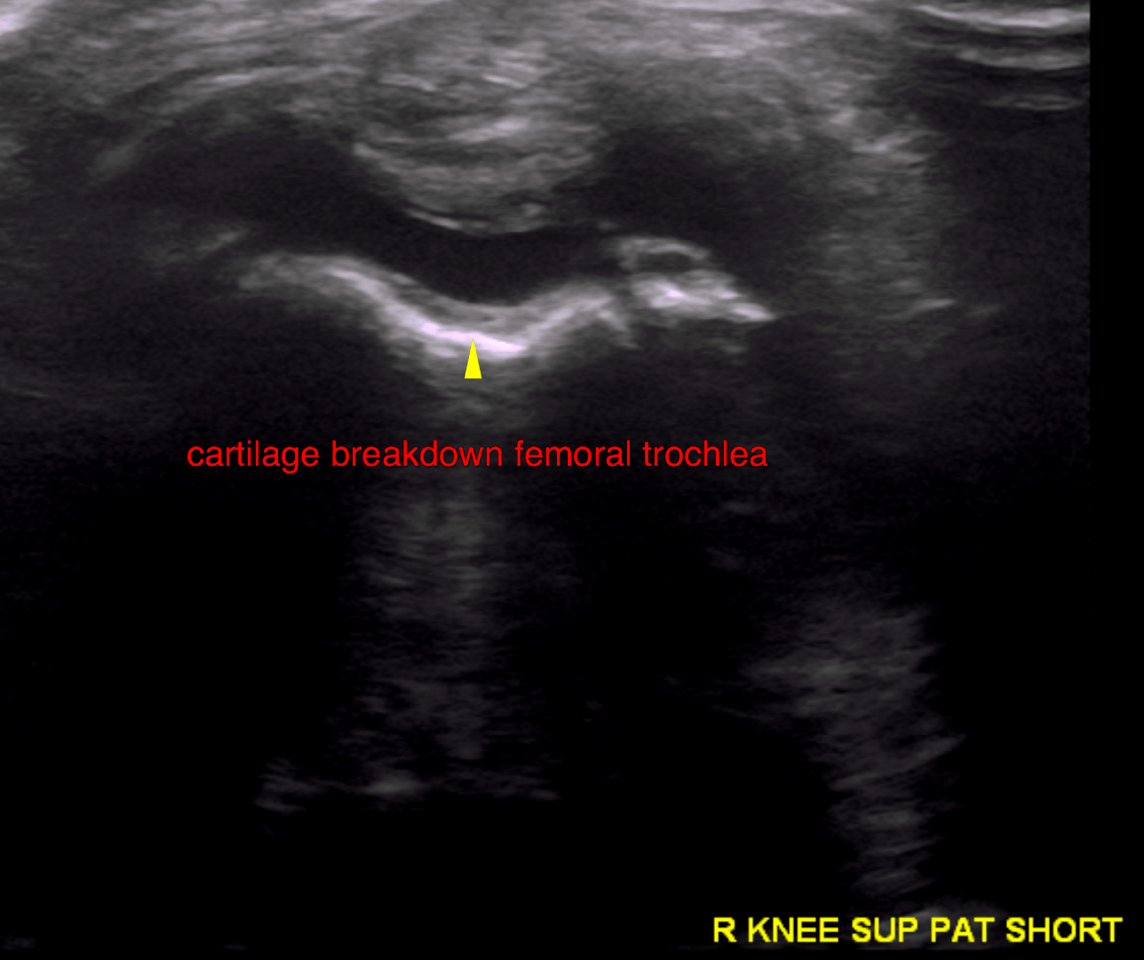
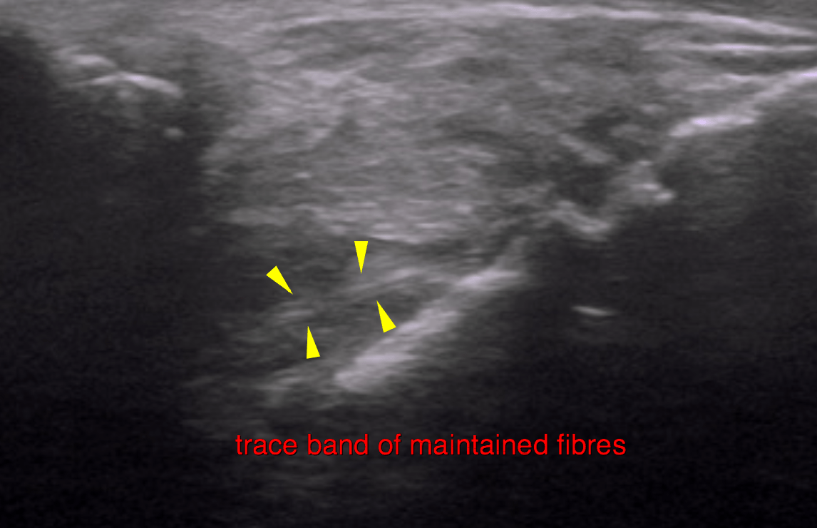
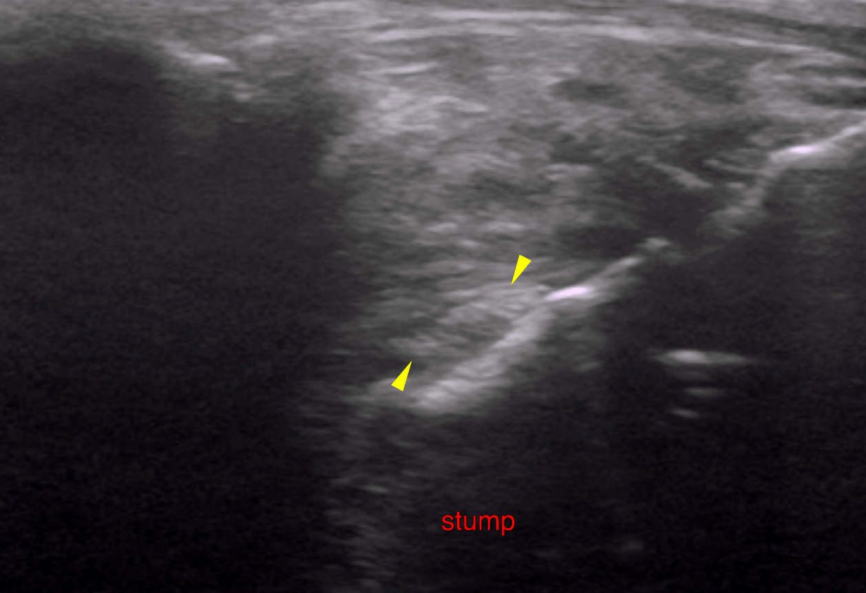
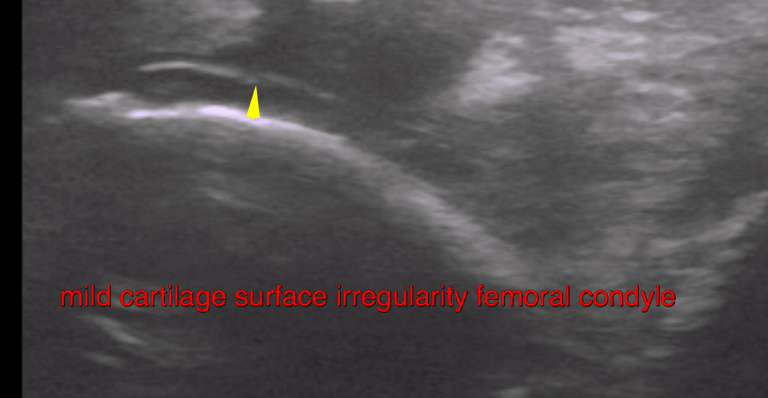
This 2 year old FS Labrador dog has a history of favoring the right hind leg for the past 6 months. Intermittent, worse after excercise and after rest.
Physical exam: positive Sits test, positive cranial drawer and CTT right stifle. Suspect CCL injury.
This 2 year old FS Labrador dog has a history of favoring the right hind leg for the past 6 months. Intermittent, worse after excercise and after rest.
Physical exam: positive Sits test, positive cranial drawer and CTT right stifle. Suspect CCL injury.