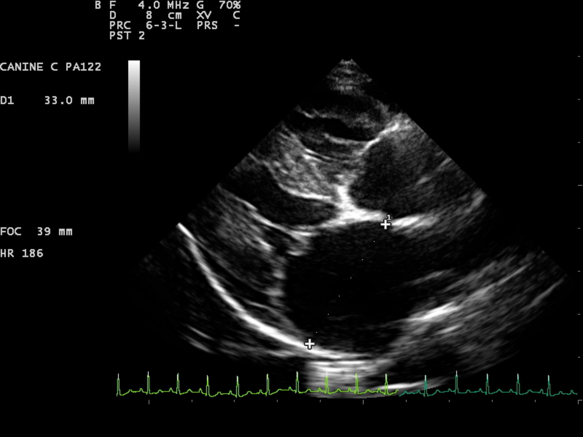
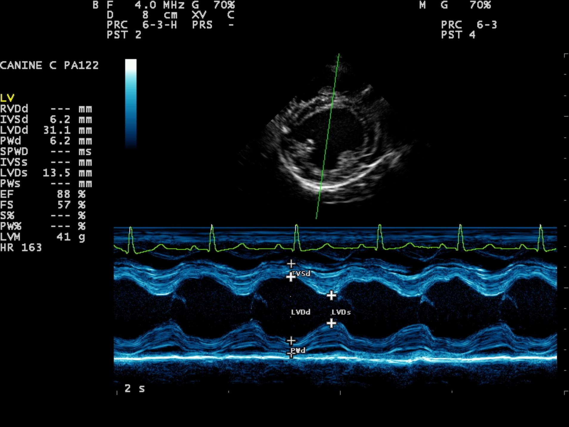
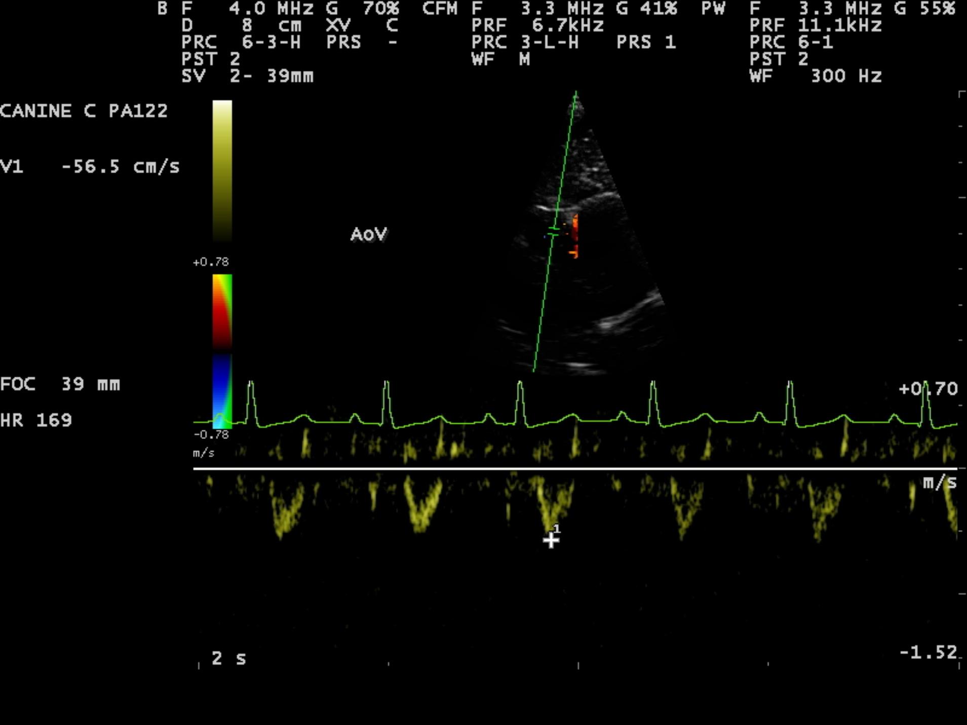
An 8-year-old F Maltese was presented for examination. A grade 5/6 systolic heart murmur was noted. Radiographs revealed severe generalized cardiomegaly and an unremarkable pulmonary parenchyma. Moderate hepatomegaly and ascites was additionally noted.
An 8-year-old F Maltese was presented for examination. A grade 5/6 systolic heart murmur was noted. Radiographs revealed severe generalized cardiomegaly and an unremarkable pulmonary parenchyma. Moderate hepatomegaly and ascites was additionally noted.