The left liver revealed a hyperechoic mass that measured 3.34 cm. This is consistent with pronounced, nodular hyperplasia or possible hepatocellular carcinoma. A separate mass that measured 4.78 x 3.4 cm with loss of detail and hyperechoic parenchyma was noted. Aside from the masses, the liver revealed swollen, irregular contour and nodular changes. Gallbladder was unremarkable. The common bile duct was unremarkable.
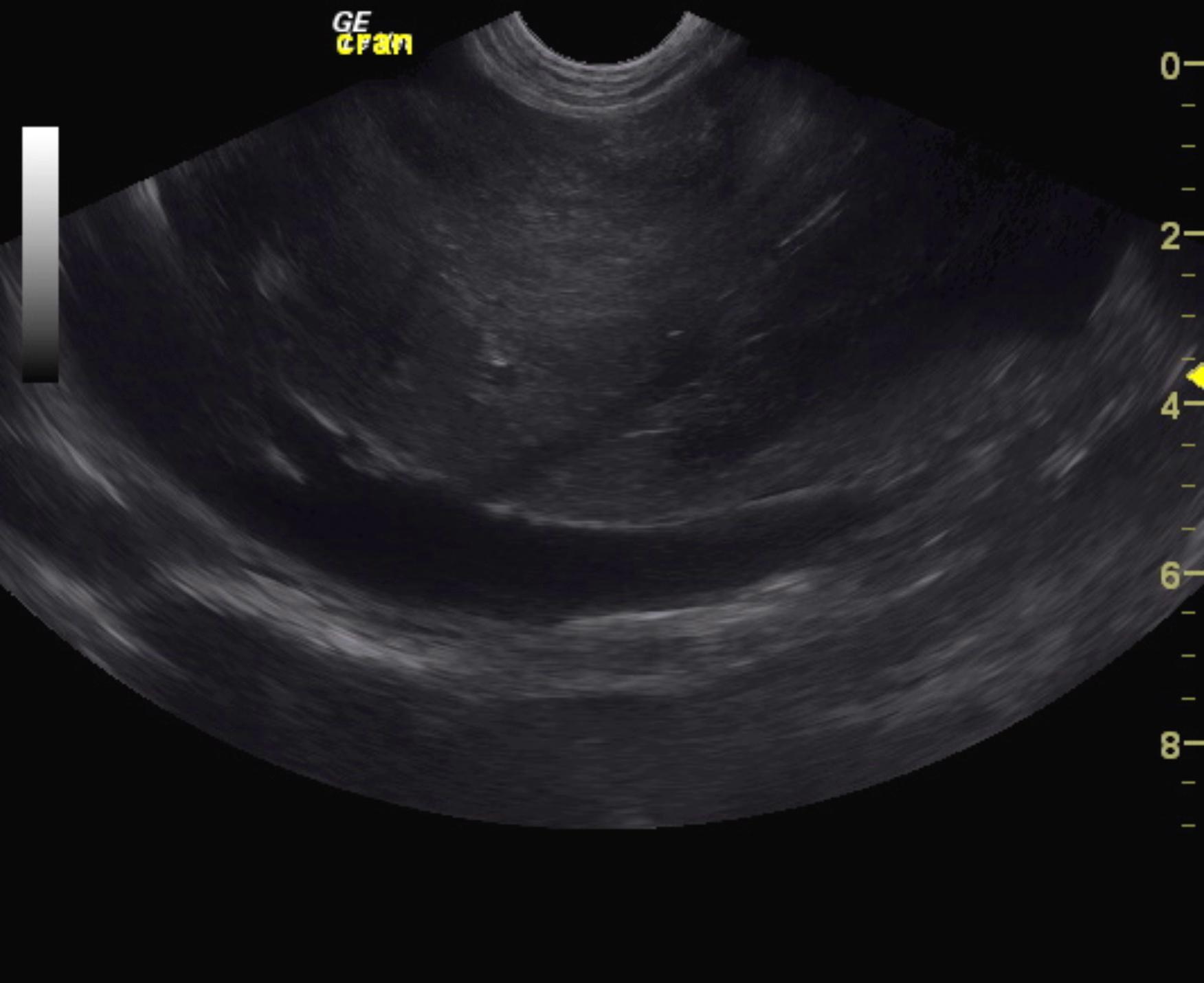
Heterogenous parenchymal changes were noted throughout the pancreas. This is likely owing to edema and artifact owing to the ascites. The abdomen presented a moderate amount of ascites, dilated hepatic veins and dilated vena cava.
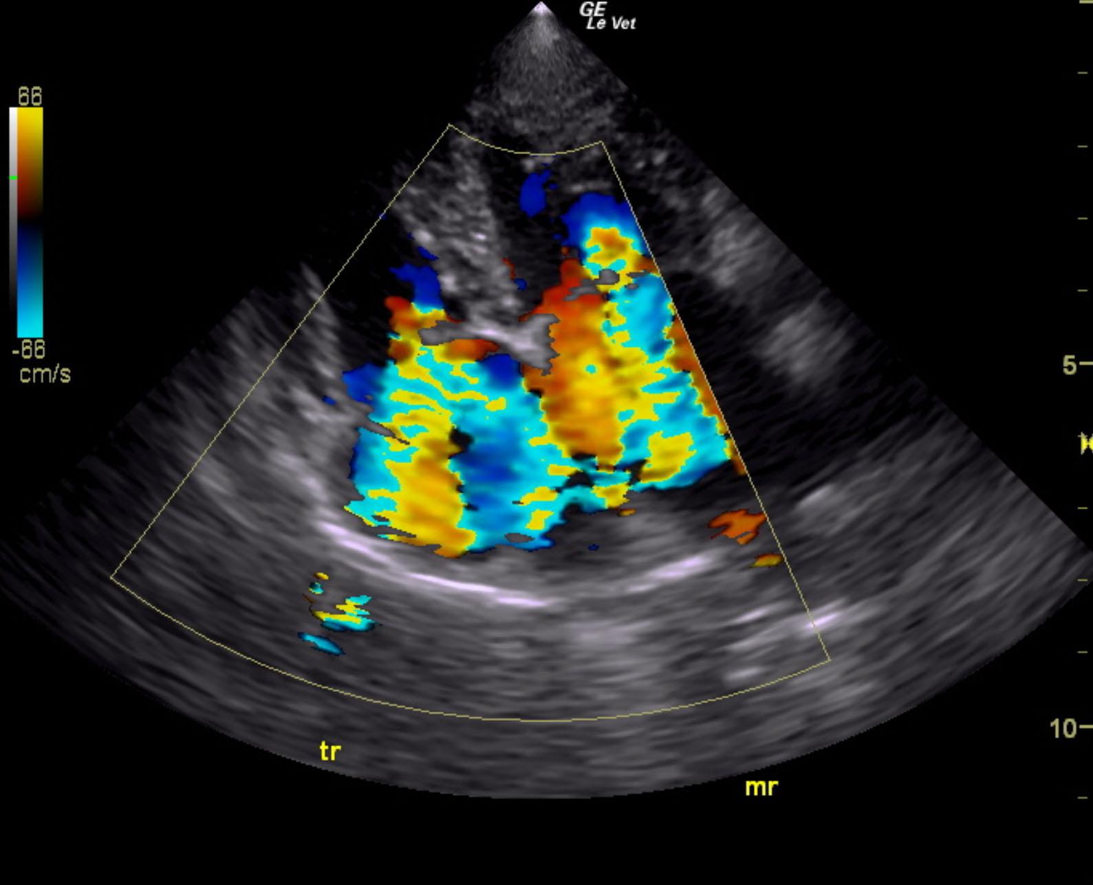
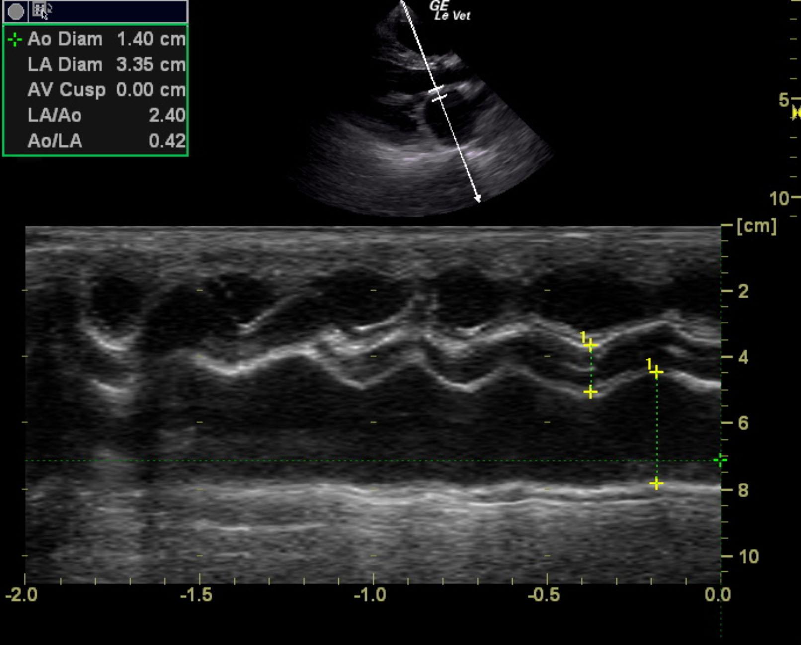
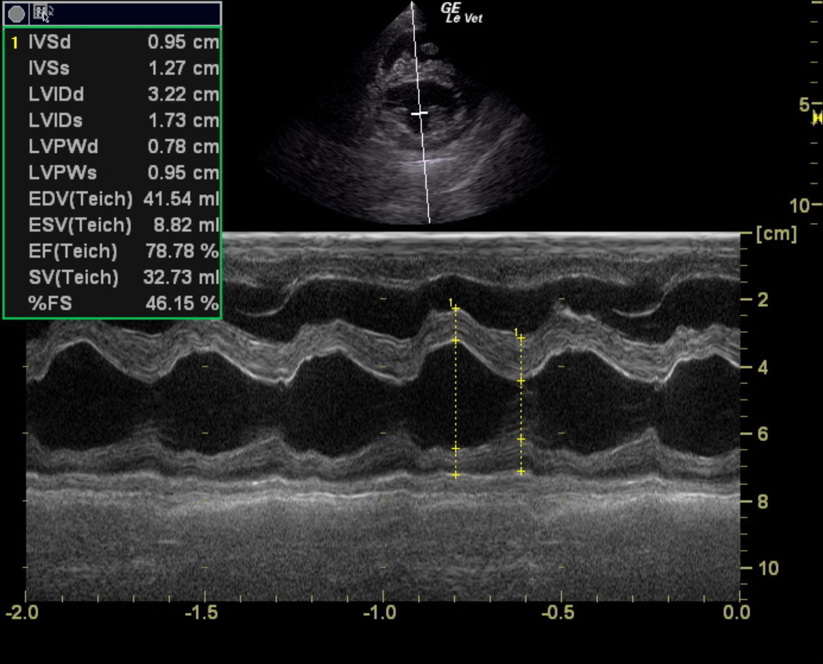
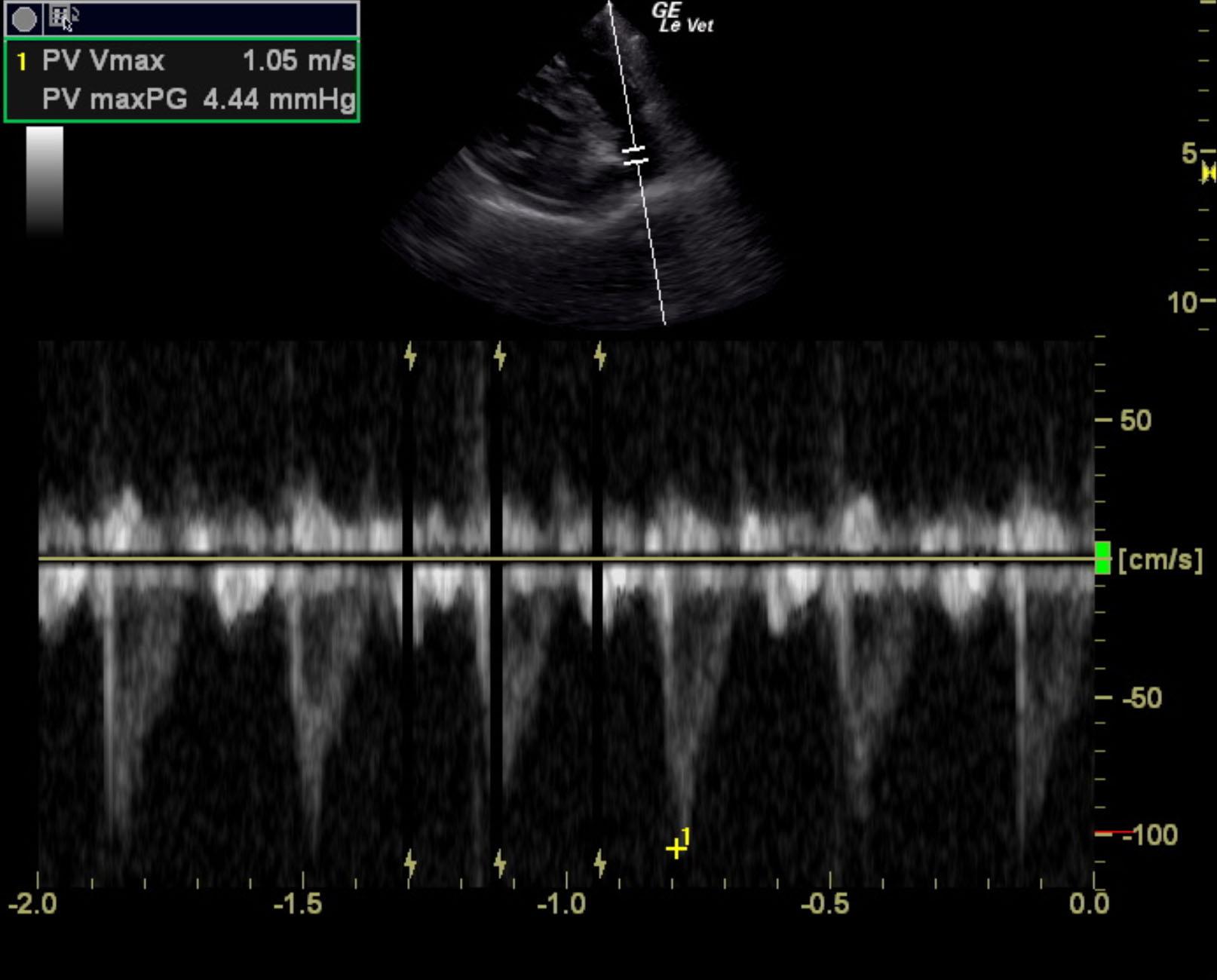
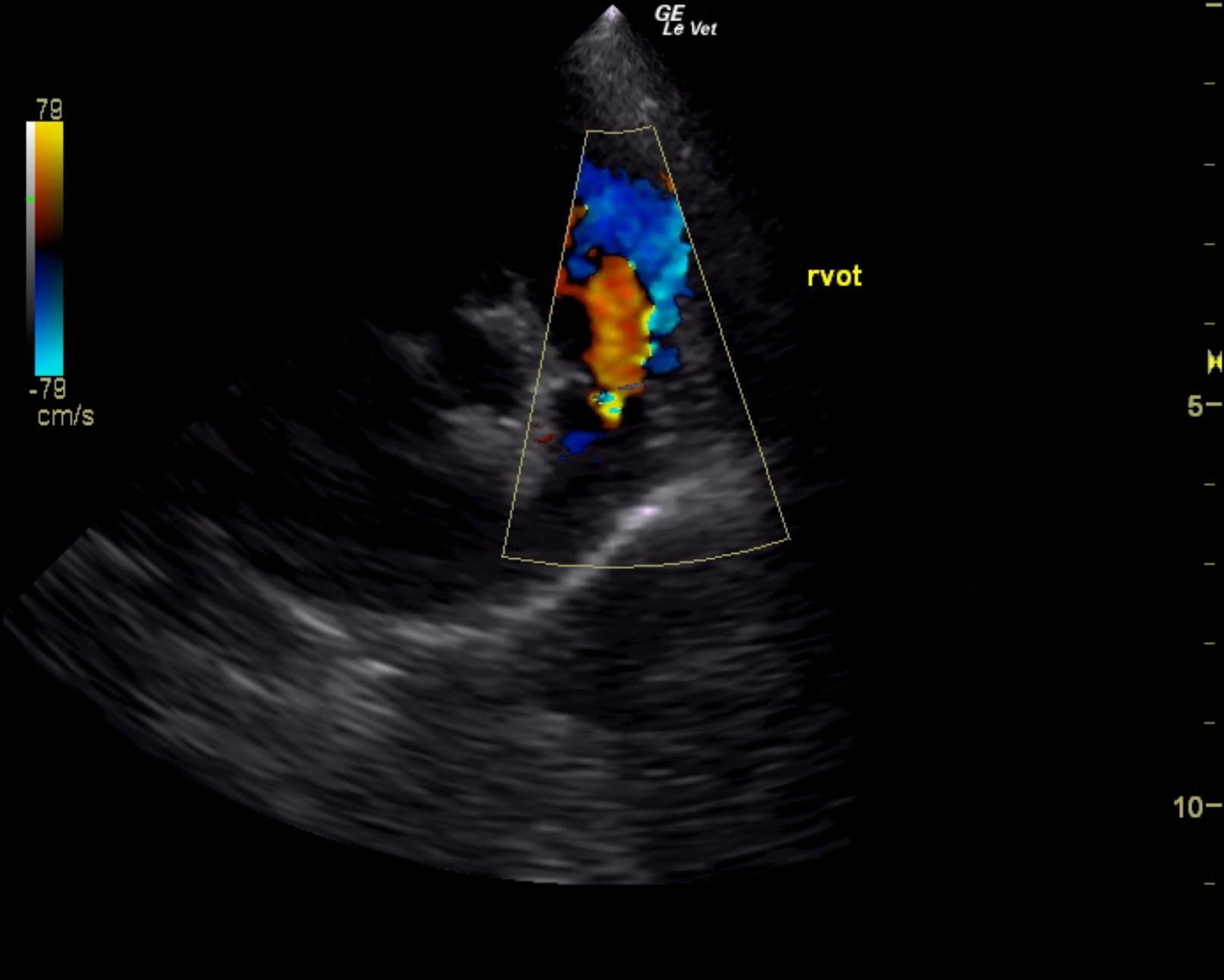
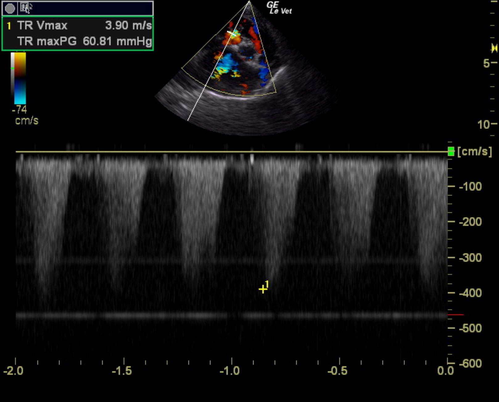
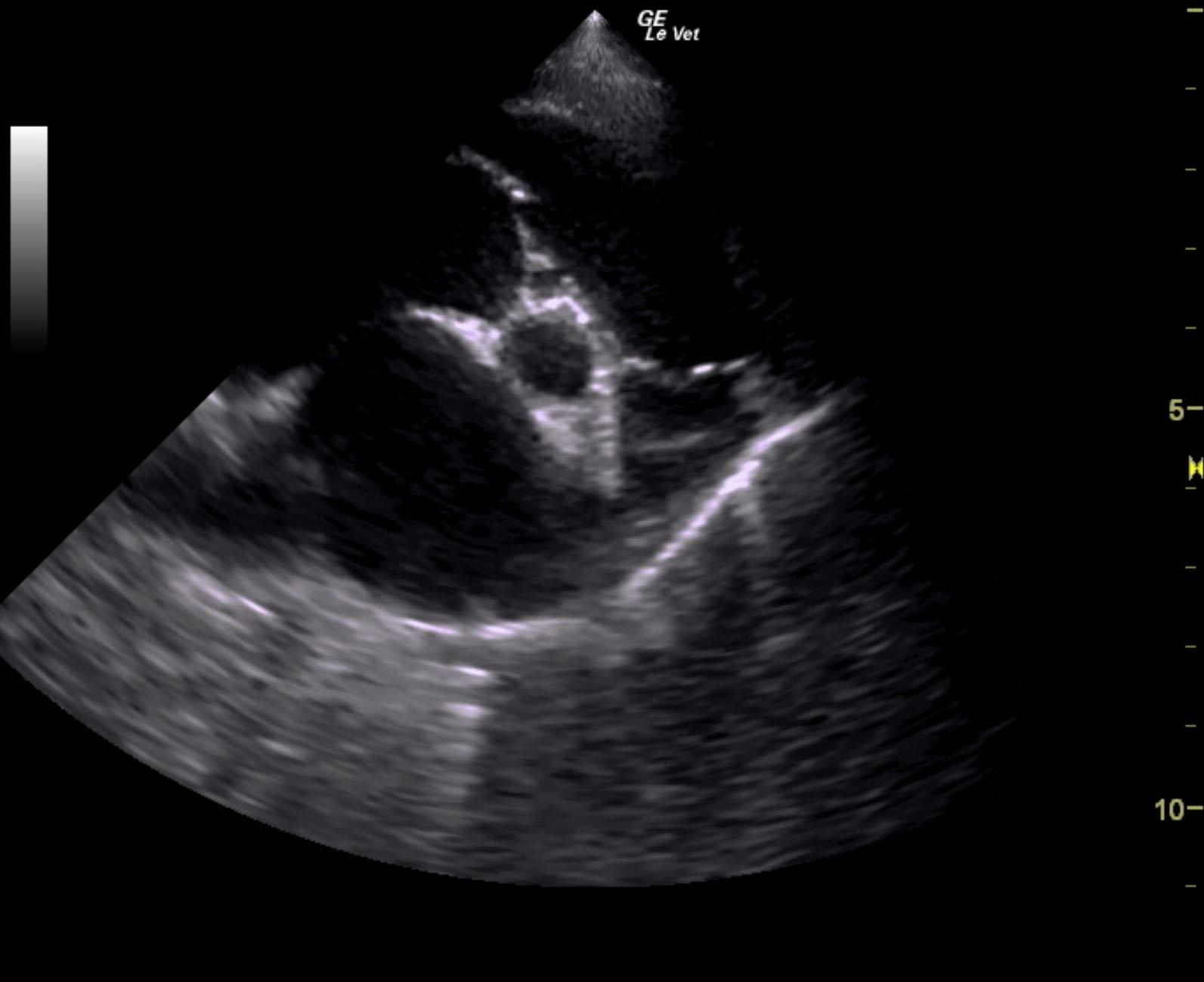
The cardiac presentation revealed moderate left and right sided overload with tricuspid and mitral valve vegetative lesions. Left atrial and right atrial enlargement was noted. Right ventricular overload was noted with flattening of the ventricular septum. Slight prolapse of the anterior mitral valve leaflet was noted. Prolapse of the tricuspid vavle was also noted. Significant pulmonic insufficiency and tricuspid insufficiency was noted. This is consistent with pulmonary hypertension.
Pulmonic insufficiency velocity 1.8 m.sec.
Tricuspid insufficiency velocity 3.9 m/sec