Miliary Lung Pattern with Peribronchial Cuffing, and Soft Tissue Lesion, consider Neoplasia in a 13 year old Chinese Crested dog

Case Study

Miliary Lung Pattern with Peribronchial Cuffing, and Soft Tissue Lesion, consider Neoplasia in a 13 year old Chinese Crested dog

History: Several months ago had an acute episode of form pitting edema that progressed to subcutaneous hemorrhage within 24 hours. Resolved within a few days. Treated with steroid injection. Tick serology was negative. Clotting panel normal except for elevated fibrinogen. Serumchemistry normal except for low creatinine. Yesterday developed acute swelling of left hind leg that progressed to subcutaneous hemorrhage today. She is experiencing tachypnea, tongue is pink.

Physical Exam: NSF except for tachypnea.

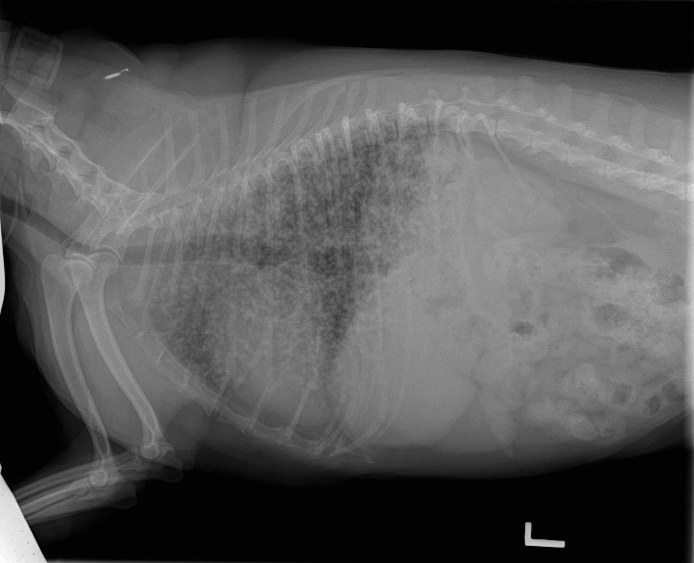
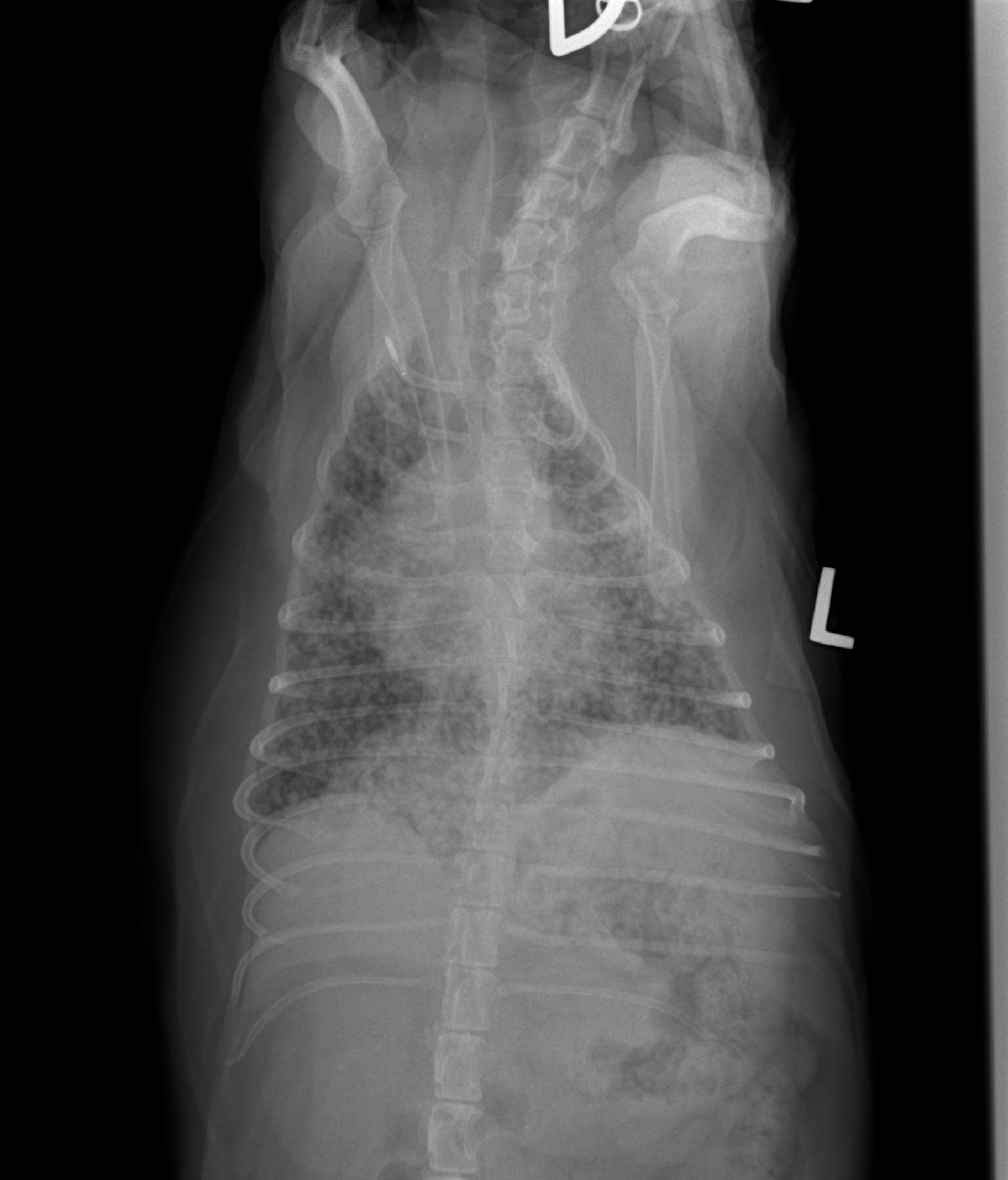
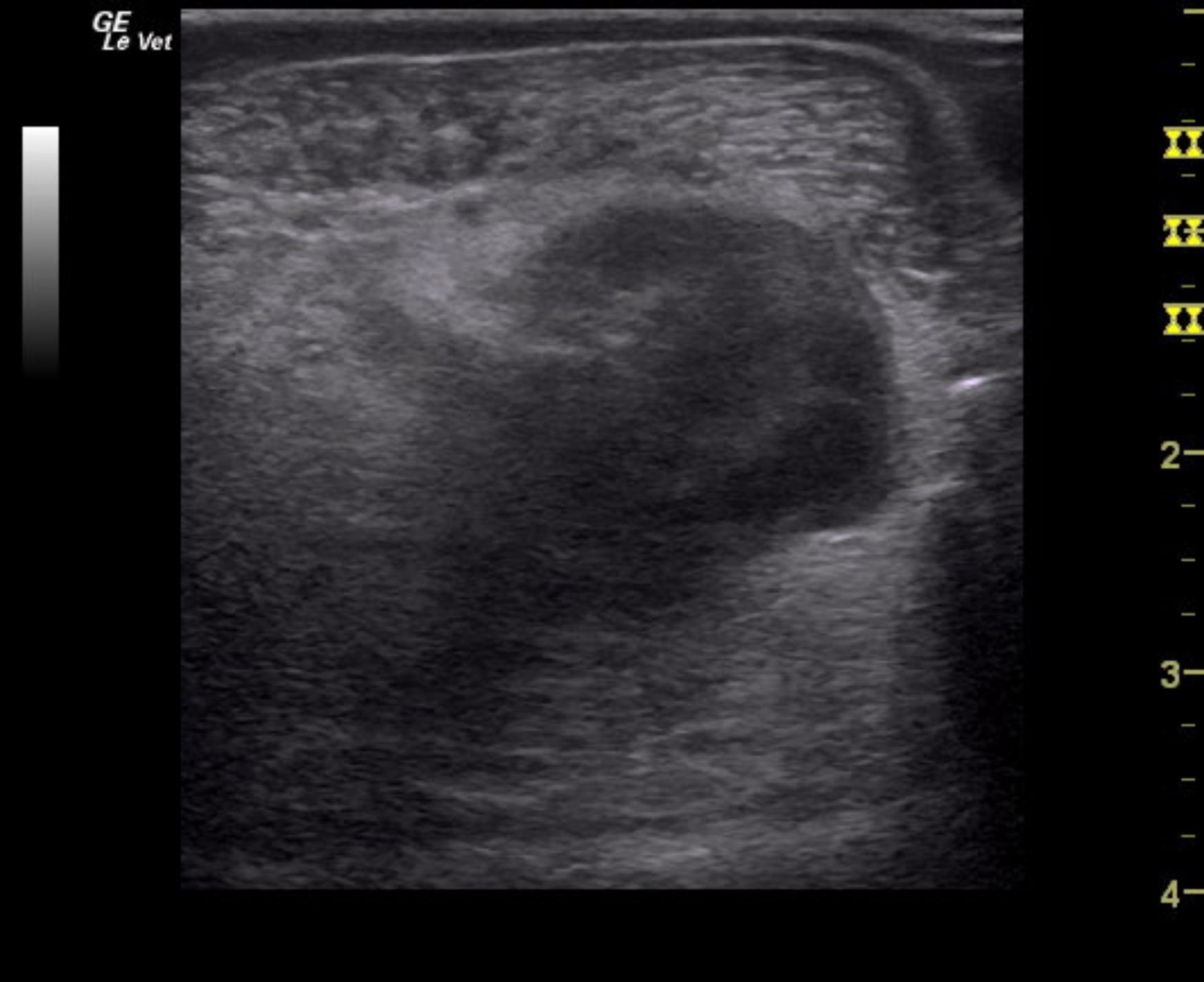
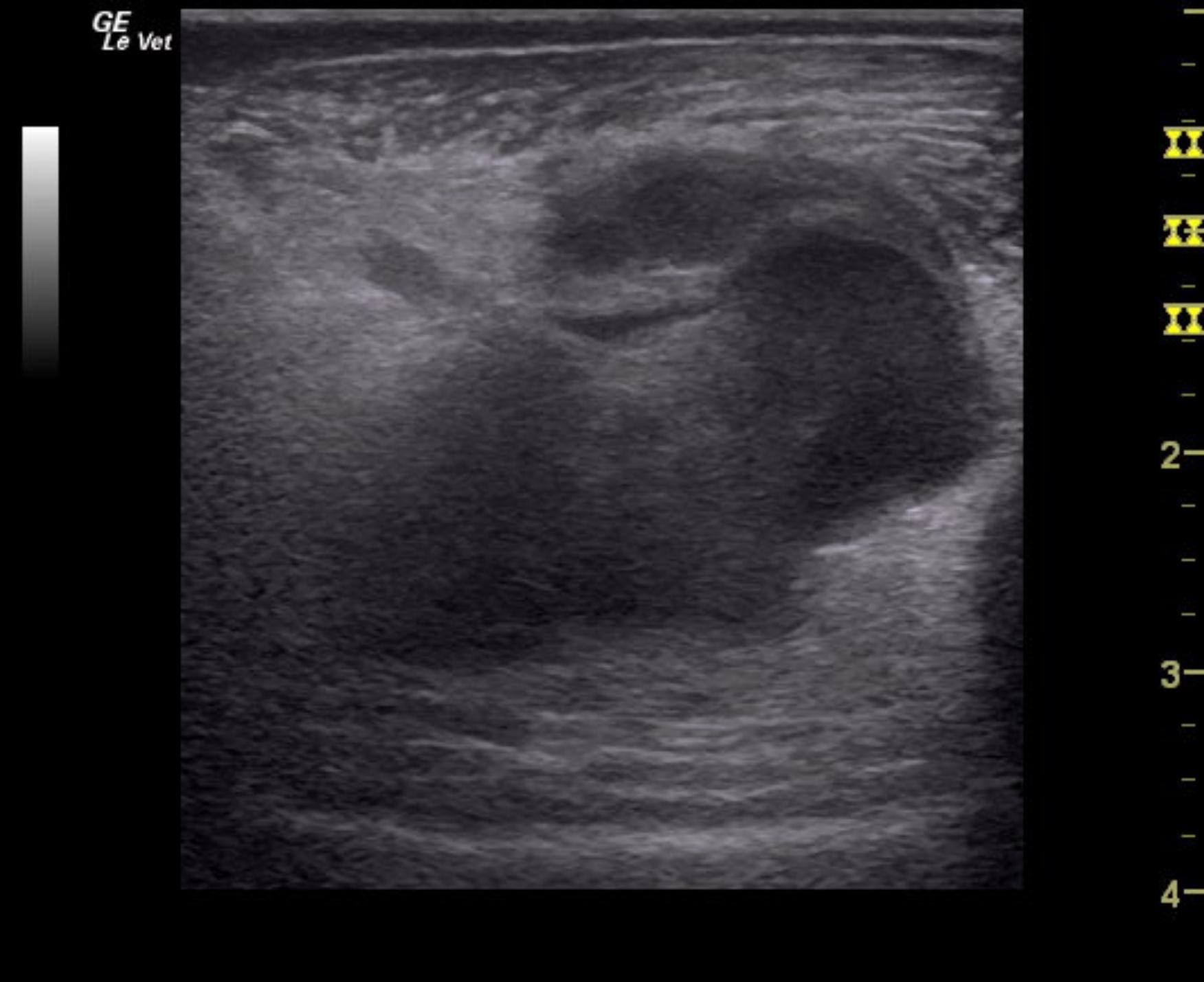
Reason for Ultrasound Exam: Very significant diffuse military lung pattern. 

Image Interpretation

Right lateral and VD thorax- Osseous structures: There was spondylosis deformans within the cranial lumbar spine.
Extrathoracic soft tissue structures: The liver margins were mildly blunted in general. One soft tissue
opaque ovoid structure compatible with a distended gallbladder or an enlarged liver lobe was seen
protruding caudoventrally from the liver margins. Both kidneys were regular in size and shape, but
showed multifocal mineral opaque structures within the region of the renal diverticuli.
Intrathoracic structures: The overall opacity of the lung was increased. There was severe generalized
bronchial/peribronchial cuffing and a miliary pattern throughout the lung. No tracheobronchial/mediastinal lymph node enlargement was seen.
A redundant tracheal membrane was seen within the caudal nach region and at the thoracic inlet. No
tracheal displacement was noted. The mediastinum was normal in width. The cardiac silhouette was
mildly obscured by the lung pattern but the heart and major vessels appeared to be within normal
limits
Ultrasound of affected limb- The ultrasonographic examination revealed an ovoid organized complex hypoechoic mass lesion centered around a misshapen aneurysmatic vessel formation. The mass measured approximately 2.5 cm in diameter. There was a significant mass effect noted displacing the surrounding anatomy and the central vessels. The adjacent soft tissues including the fascial planes and muscles presented moderate generalized swelling and hyperemic inflammatory edema. Inter- and subfascial anechoic areas lacking vascularization were seen and compatible with fresh hemorrhage.
In one of the video loops a superficial lymph node regular in size, shape and echoarchitecture was seen.

DX

The radiographs show a severe miliary lung pattern with peribronchial cuffing. Neoplastic infiltration such as primary bronchoalveolar carcinoma, lymphoma, or less likely granulomatous lymphomatosis and histiocytic sarcoma have to be considered. The ultrasonographic findings are indicative of a soft tissue mass lesion meeting neoplastic criteria. Differential diagnoses include a round cell neoplasia such as lymphosarcoma, histiocytic sarcoma or mast cell tumor as well as a hemangiosarcoma/-endothelioma or other.RADS

Comments

RADs – The main differential diagnoses in this case are eosinophilic bronchopneumopathy (formerly pulmonary infiltrates with eosinophils) versus granulomatous pulmonary disease due to fungal (depending on endemic region) or other opportunistic infection.

The findings are not typical for ARDS or pulmonary hemorrhage – this is usually predominantly alveolar when severe – can be uniform or patchy, but not bronchial/miliary. A bronchoscopy with deep bronchoalveolar lavage and/or fine needle aspiration of the lung may be considered for further workup.

Ultrasound- 

The potential for a simple aneurysm and hematoma is low especially taking the radiographically diagnosed lung changes into account. There is secondary hemorrhage and inflammatory limb edema which is inter- and subfascial in distribution. There also is potential for development of a compartment syndrome, so the patient needs clinical monitoring for subfascial pressure increase. 

Further work up requires sampling of the mass lesion. Ultrasound guided Fine needle aspiration avoiding the central vessels is recommended as there is potential for a mast cell or round cell neoplasia. Proactive antihistamic treatment should be enforced before sampling. 


Clinical Differential Diagnosis

DDx= metastatic neoplasia, embolic process, vasculitis, bronchopneumonia, allergic bronchitis, fungal infection. Determine if there is ultrasound evidence of disease that could be causing this reaction in lungs.

Patient Information

Patient Name : Willow Cerf, Ridgewood AH
Gender : Female, Spayed
Species : Canine
Type of Imaging : Ultrasound
Status : Complete
Liz Wuz Here : Yes

Clinical Signs

  • Swollen limb

Exam Finding

  • Swollen limb
  • Tachypnea

Images

rotated11willow_cerf_leg_mass_met_lung_patternwillow_cerf_met_lung_pattern_leg_mass_2

Clinical Signs

  • Swollen limb
Skip to content