Lymphoma in a 3 year old FS Rhodesian Ridgeback

Case Study

Lymphoma in a 3 year old FS Rhodesian Ridgeback

A 3-year-old FS Rhodesian Ridgeback was presented for the evaluation of enlarged lymph nodes of 3 weeks’ duration. Generalized lymphadenopathy was present on physical examination. The serum chemistry showed hyperglobulinemia, and thrombocytopenia was present on the CBC. Thoracic radiographs did not show any signs of lymphadenopathy or any lesions consistent with neoplasia.
A 3-year-old FS Rhodesian Ridgeback was presented for the evaluation of enlarged lymph nodes of 3 weeks’ duration. Generalized lymphadenopathy was present on physical examination. The serum chemistry showed hyperglobulinemia, and thrombocytopenia was present on the CBC. Thoracic radiographs did not show any signs of lymphadenopathy or any lesions consistent with neoplasia.

Sonographic Differential Diagnosis

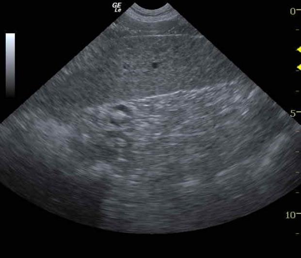
The sonographic appearance is consistent with infilitrative and multicentric lymphoma or similar neoplasia.

Image Interpretation

The spleen was mildly enlarged and appeared moth-eaten, which is consistent with infiltrative disease. The liver also demonstrated micronodular changes and periportal lymphadenopathy.

DX

Lymphoma

Outcome

The patient was discharged with a course of antibiotics pending the cytology results.

Comments

This a presumptive diagnosis of splenic and multicentric lymphoma based on the LN aspirates. The “moth eaten” splenic presentation with multicentric lymphadenopathy is consistent with this diagnosis. Technically, splenic and abdominal LN aspirates would have been appropriate but were declined by the owner as it would not have changed the chemotherapeutic protocol. This was an ultrasound performed for staging and evaluation of infiltrative patterns in the visceral organs.

Clinical Differential Diagnosis

Malignant lymphoma. Thrombocytopenia – lymphoma, infectious disease (especially rickettsia disease such as salmon poisoning or Ehrlichia), leptospirosis.

Sampling

US guided FNAs. Cytology results from the prescapular and popliteal lymph nodes were consistent with a diagnosis of malignant lymphoma. The Madison-Wisconsin chemotherapeutic protocol was initiated.

Patient Information

Patient Name : Brodie M
Gender : Female, Spayed
Species : Canine
Type of Imaging : Ultrasound
Status : Complete
Liz Wuz Here : Yes
Code : 08_00011

Clinical Signs

  • Enlarged Lymph Nodes

Exam Finding

  • Enlarged Lymph Nodes

Images

spleen

Blood Chemistry

  • Globulin, High

CBC

  • Platelet Count, Low

Clinical Signs

  • Enlarged Lymph Nodes