An 8-year-old FS large mixed breed dog presented for lethargy and anorexia. Weight loss was found on an otherwise normal physical examination. Hepatic enzymes were normal on initial presentation, but hypoalbuminemia was present. A follow-up blood chemistry profile revealed a moderate hypoalbuminemia and slightly increased SAP and slightly increased ALT levels. A mild to moderate neutrophilic monocytic leukocytosis also developed over a 3-week period. Mildly prolonged PT and aPTT were apparent at the time of the sonogram. Urinalysis was unavailable at the time of the sonogram.
An 8-year-old FS large mixed breed dog presented for lethargy and anorexia. Weight loss was found on an otherwise normal physical examination. Hepatic enzymes were normal on initial presentation, but hypoalbuminemia was present. A follow-up blood chemistry profile revealed a moderate hypoalbuminemia and slightly increased SAP and slightly increased ALT levels. A mild to moderate neutrophilic monocytic leukocytosis also developed over a 3-week period. Mildly prolonged PT and aPTT were apparent at the time of the sonogram. Urinalysis was unavailable at the time of the sonogram.
Case Study
Lymphohistiocytic neutrophilic hepatitis, portal hypertension, copper accumulation in a 8 year old FS mixed breed dog
Sonographic Differential Diagnosis
Chronic inflammatory hepatopathy with suggested fibrosis and/or hyperplasia. Neoplasia also could be considered.
Image Interpretation
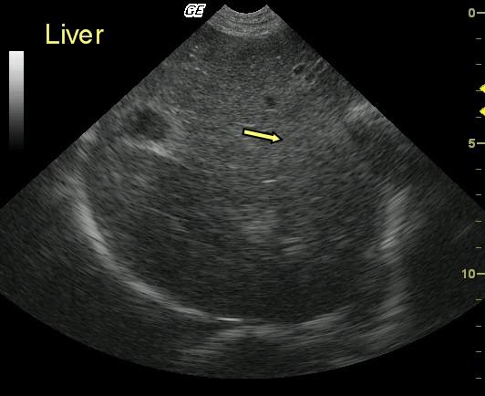
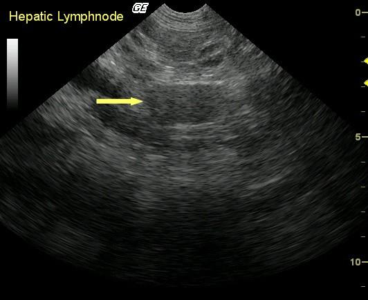
The sonogram reveals a mild, diffusely heteroechoic hepatic echotexture with multiple ill-defined, irregularly shaped echogenic foci (arrow). Periportal lymphadenopathy was present as well.
DX
Outcome
The patient is responding well to a broad-spectrum hepatic support protocol including zinc sulfate. Repeat biopsy for further copper assessment and progressive structural changes in the future ideally would have been warranted here.
Comments
This case demonstrates the importance of large core biopsy samples in cases of periportal lymphadenopathy that can represent, but is surely not pathognomonic for, infiltrative neoplasia. A moderately aggressive hepatitis with mild portal hypertension was this patient’s underlying disease
Clinical Differential Diagnosis
Liver pathology: Hepatitis, neoplasia, protein-losing enteropathy, protein-losing nephropathy, biliary calculi, gallbladder mucocele.
Sampling
Two 16-gauge US-guided biopsy specimens obtained after vitamin K administration revealed moderate portal and random lymphohistiocytic, neutrophilic hepatitis with mild portal fibrosis. Twenty percent of the hepatocytes revealed moderate copper accumulation suggestive of some influence of copper toxicosis. Bile duct hyperplasia and superimposed hepatocellular degeneration and cholestasis also were evident.
Video
Patient Information
Clinical Signs
- Anorexia
- Lethargy
Exam Finding
- Weight loss
Images
Blood Chemistry
- Albumin, Low
- Alkaline Phosphatase (SAP), High
- ALT (SGPT), High
CBC
- Monocytes, High
Clinical Signs
- Anorexia
- Lethargy
Special Testing
- PT Prolonged
- PTT Prolonged