An 8-year-old MN Golden Retriever was presented for a second opinion following diagnosis of possible torn cruciate ligament two weeks prior and decreased appetite. Physical examination was consistent with a probable torn cruciate and patient sedated for orthopedic radiographs. At this point it was discovered that he was febrile (103°F). Three days later he was represented for anorexia and weight loss. On physical examination the mucous membranes were slightly pale and there had been a 5-6 lbs weight loss over a two month timeframe.
An 8-year-old MN Golden Retriever was presented for a second opinion following diagnosis of possible torn cruciate ligament two weeks prior and decreased appetite. Physical examination was consistent with a probable torn cruciate and patient sedated for orthopedic radiographs. At this point it was discovered that he was febrile (103°F). Three days later he was represented for anorexia and weight loss. On physical examination the mucous membranes were slightly pale and there had been a 5-6 lbs weight loss over a two month timeframe. In-house coagulation panel was within normal limits, and platelet count was high.
Case Study
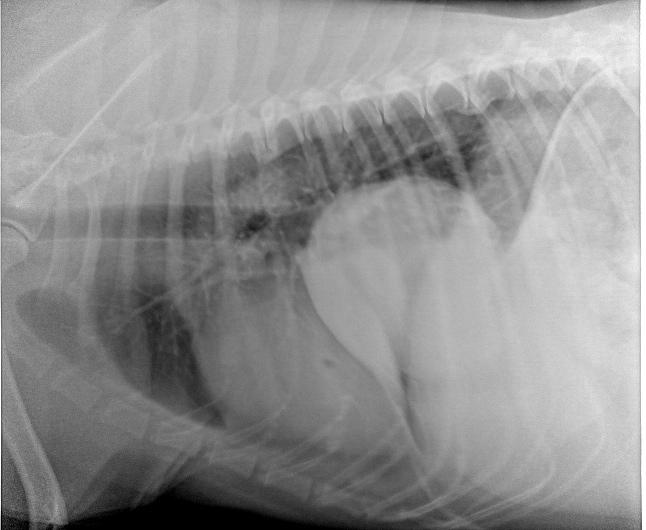
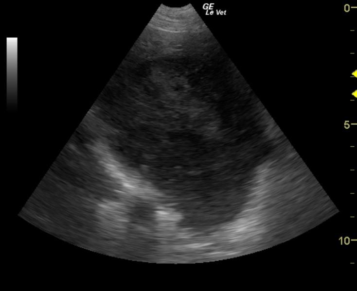
Lung mass in an 8 year old MN Golden Retriever dog
Sonographic Differential Diagnosis
Solitary lung mass. Rule outs would include sarcoma or carcinoma with a minor potential for lobar lung necrosis. This mass is likely primary lung in origin.
Image Interpretation
A mixed heterogenic and dramatically hypoechoic left caudal lung mass is present. There is air entrapment in the far field which indicates lung origin. Ultrasound-guided fine needle aspirates were performed. The lesion appears isolated to the appropriate lung lobe and may be resectable.
DX
Outcome
Fine needle aspirates taken from the left caudal lung lobe mass found evidence of chronic inflammation and mild acute hemorrhage. Fluid sampled surrounding the left lung lobe mass was negative for bacterial growth. The patient was treated with NSAIDs and antibiotics with the recommendation that he be referred to a surgeon for probable lung lobe resection with biopsy. Several weeks later under suspicion of the presence of lung cancer by owner, the patient was euthanized.
Comments
Often inflammation is present along with neoplasia. Core biopsy would have been a better approach in this particular lesion as adequate window was possible for biopsy.
Clinical Differential Diagnosis
Anemia, reduced perfusion, cardiac disease, neoplasia
Sampling
US-guided FNAs taken from the left caudal lung lobe mass found evidence of chronic inflammation and mild acute hemorrhage. Fluid sampled surrounding the left lung lobe mass was negative for bacterial growth.
Video
Patient Information
Clinical Signs
- Anorexia
- Fever
Exam Finding
- Pale Mucous Membranes
- Weight loss
Images
CBC
- Platelet Count, High
Clinical Signs
- Anorexia
- Fever