Leptospirosis suspected in a 3 year old FS French Bulldog

Case Study

Leptospirosis suspected in a 3 year old FS French Bulldog

A 3-year-old FS French Bulldog with a history of progressive elevation of liver enzyme activity was presented for evaluation of lethargy and vomiting. On urinalysis inappropriate SG, proteinuria, bilirubinuria, and hematuria were present. Abnormalities on serum biochemistry were prolonged PT, elevated ALP activity and bilirubin, and severely elevated ALT activity.
A 3-year-old FS French Bulldog with a history of progressive elevation of liver enzyme activity was presented for evaluation of lethargy and vomiting. On urinalysis inappropriate SG, proteinuria, bilirubinuria, and hematuria were present. Abnormalities on serum biochemistry were prolonged PT, elevated ALP activity and bilirubin, and severely elevated ALT activity.

Sonographic Differential Diagnosis

Acute nephritis, hepatitis presentation. Concurrent microvascular dysplasia, portal vein hypoplasia is possible. However, this is not an issue in this case. I strongly suspect Leptospirosis.
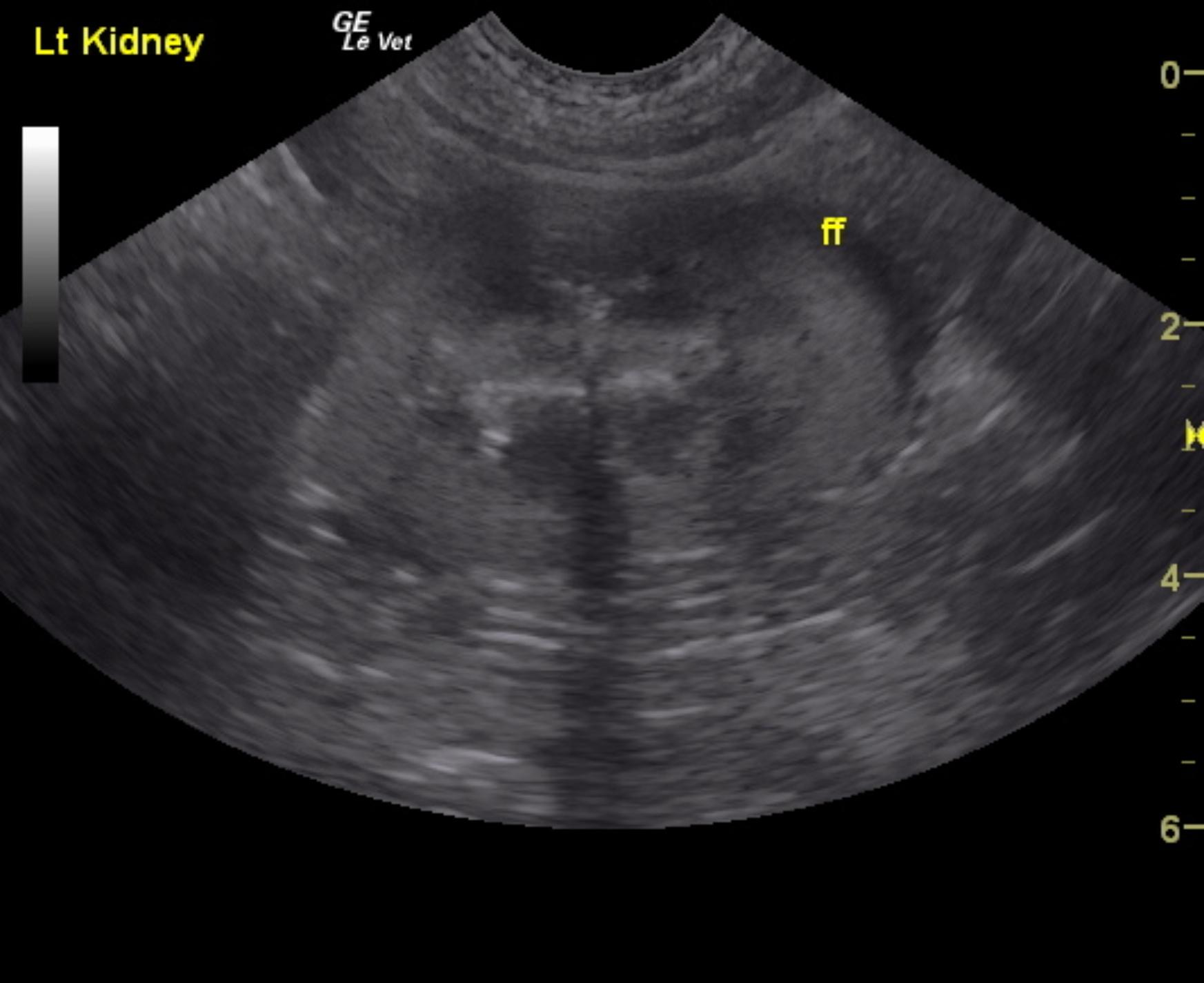
Given the clinical profile and the free fluid around the kidneys this is a classic presentation for acute Leptospirosis especially given that the abdomen is structurally unremarkable other than the free fluid and mild microhepatica which is not a clinical issue at this point.

Image Interpretation

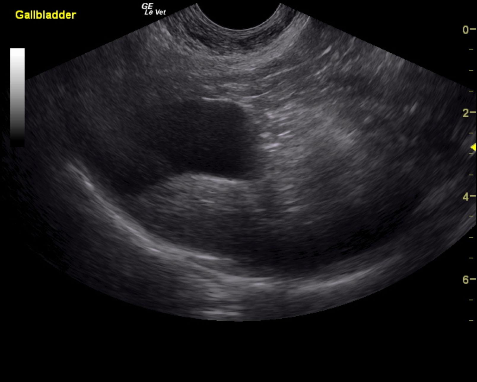
The kidneys revealed normal size and structure, corticomedullary definition and ratio (cortex 1/3 of medulla). The cortices presented largely uniform texture with normal echogenic relationship to liver and spleen. Medullary echogenicity differed distinctly from that of the cortex and no evidence of dilation could be seen. The capsules were acceptably uniform without dramatic irregularities. The left kidney measured 5.02 cm. The right kidney measured 5.42 cm. A slight amount of free fluid was noted caudal to the kidneys. This is consistent with acute nephritis. The liver was subnormal in size, yet structurally unremarkable. Portal vein, vena cava and aortic ratio was 1:1:1. No evidence of intrahepatic or extrahepatic shunts. The gallbladder was unremarkable.

DX

Suspect Leptospirosis

Outcome

None

Comments

Recommend plasma transfusion, IV Ampicillin, gastrointestinal protectants and hepatic support in this patient. Doxycycline is also warranted. Reassessment of the liver enzymes is recommended over the next 48-72 hours. Guarded to good prognosis assuming this is Leptospirosis. Other acute hepatic renal insults are possible.

Clinical Differential Diagnosis

Liver pathology– acute hepatitis (viral, bacterial, toxins), chronic-active hepatitis, abscessation, granulomatous disease
Gall bladder – cholecystitis
Renal pathology – acute kidney injury, pyelonephritis
Leptospirosis

Sampling

None

Patient Information

Patient Name : Colvin I
Gender : Female, Spayed
Species : Canine
Type of Imaging : Ultrasound
Status : Complete
Liz Wuz Here : Yes
Code : 06_00183

Clinical Signs

  • Lethargy
  • Vomiting

Images

lepto_kidney_free_fluidcolvin_ingoglia_lepto_liver

Blood Chemistry

  • Alkaline Phosphatase (SAP), High
  • ALT (SGPT), High
  • Total Bilirubin, High

Clinical Signs

  • Lethargy
  • Vomiting

Special Testing

  • PT Prolonged

Urinalysi

  • Bilirubin Present
  • Blood Present
  • Protein Present
  • Specific Gravity Abnormal