A 9 year old MN Maltese dog was presented for evaluation of progressive anorexia and soft feces. Physical examination and rectal palpation were both within normal limits. Fecal and urinalysis, CBC, serum biochemistry, cPl, and survey radiographs of the thorax and abdomen were all within normal limits.
A 9 year old MN Maltese dog was presented for evaluation of progressive anorexia and soft feces. Physical examination and rectal palpation were both within normal limits. Fecal and urinalysis, CBC, serum biochemistry, cPl, and survey radiographs of the thorax and abdomen were all within normal limits.
Case Study
Lacteal Dilatation 9 Year Old MN Maltese dog
Sonographic Differential Diagnosis
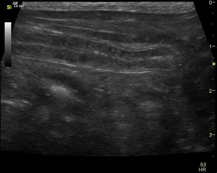
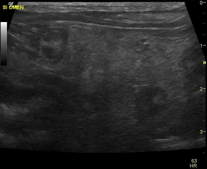
Necrotic portion of small intestine with reactive omentum and mucosal striations.
Image Interpretation
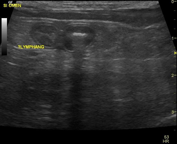
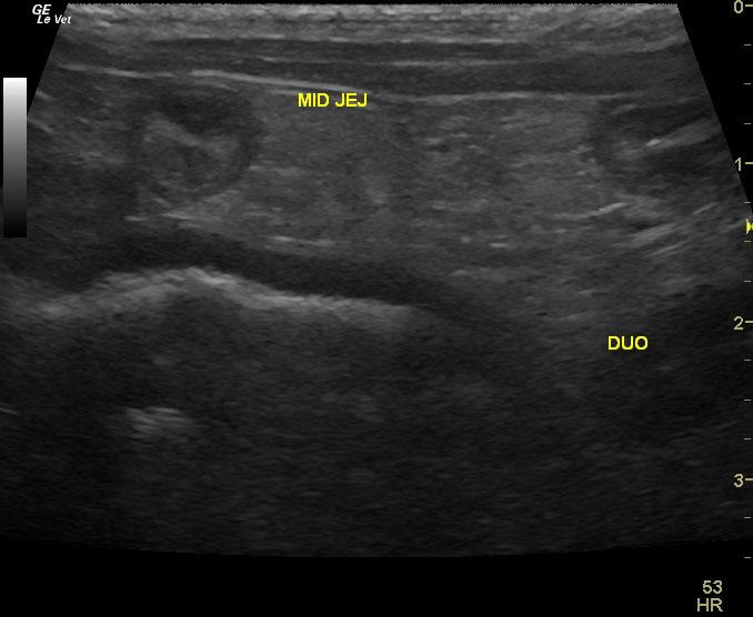
The gastrointestinal tract revealed areas of mucosal striations in the small intestine with surrounding echogenic omentum. A large region of the small intestine appeared involved and measured approximately 8.0 cm in length. This region of the intestinal tract revealed numerous mucosal striations. Spontaneous intestinal necrosis is likely the underlying cause in this case. This is suggestive for both inflammation and potential lymphangectasia. Strongly recommend removal of this area of intestine. Intraoperative ultrasound would be ideal in this case.
DX
Exploratory surgery revealed dilated lacteals in duodenum and jejunum, thickened muscularis.
Outcome
On prednisone and Pepcid post surgical. The patient is eating and doing much better.
Comments
Strongly recommend intraoperative ultrasound and intestinal resection in this patient. The sonographer may be able to identify the areas of the healthiest bowel in order to resect and anastamosis the borders of the unhealthy intestine. The primary differentials for this portion of intestine would be chronic inflammatory bowel disease with transmural inflammation and likely lymphangectasia. Omentopexy would likely be recommended post anastamosis. Alternatively, the patient could be treated with 72 hour IV fluid protocol and broad spectrum antibiotics in order to combat any transmural infection that may have occurred with bacteria deriving from the lumen that may be complicating this presentation. However, the bowel appears chronically unhealthy and therefore, ideally would be resected. If intraoperative ultrasound is not possible then the surgeon may not see the entire portion of affected small intestine. Landmarks would include any areas of reactive omentum associated with small intestine and resection 10 cm on either side of reactive omentum; however, this would be less precise than with intraoperative ultrasound guided surgery.
Clinical Differential Diagnosis
GI tract – neoplasia, IBD, foreign body, granulomatous enteritis, intussusception, focal perforation
Sampling
none was performed
Video
Patient Information
Patient Name :
Lucky H
Gender :
Male, Neutered
Species :
Canine
Type of Imaging : Ultrasound
Status :
Complete
Liz Wuz Here :
Yes
Code :
04_00321
Clinical Signs
- Anorexia
- Soft stool
Images
Clinical Signs
- Anorexia
- Soft stool