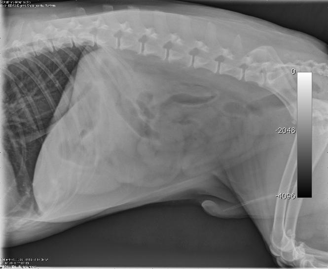
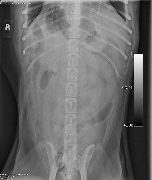
A 10 year-old MN Shepherd mixed dog presented with a 2-3 month history of intermittent vomiting and a more recent anorexia. Abnormalities on physical examination were dehydration and a 3 cm firm left anal sac mass. The only significant finding on CBC and serum biochemistry was hypoalbuminemia. Survey radiographs showed no evidence of pulmonary metastases but revealed excessive pyloric gas pattern with a volume contracted heart.
A 10 year-old MN Shepherd mixed dog presented with a 2-3 month history of intermittent vomiting and a more recent anorexia. Abnormalities on physical examination were dehydration and a 3 cm firm left anal sac mass. The only significant finding on CBC and serum biochemistry was hypoalbuminemia. Survey radiographs showed no evidence of pulmonary metastases but revealed excessive pyloric gas pattern with a volume contracted heart.
Case Study
Intussusception in a 10 year old MN German Shepherd mixed dog
Sonographic Differential Diagnosis
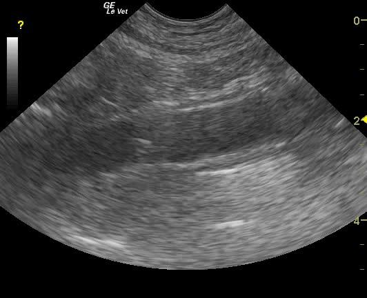
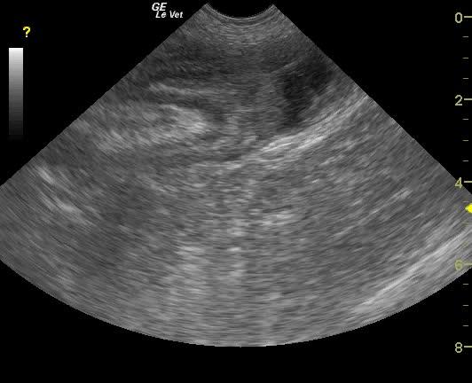
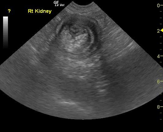
Ileocecocolic intussusception. Moderate chronic renal disease.
Image Interpretation
The distal small intestine in this patient presented an obstructive pattern with dilated small intestine followed by what appeared to be ileocecocolic intussusception. This was confirmed in long and short axis. Some hyperechoic reactive fat was present within the intussusception itself as well as associated with the serosa.
DX
Outcome
The patient was taken to surgery and a large 15 cm intussusception was found along the distal jejunum, which could not be reduced during surgery. There was a 3 cm firm intraluminal mass causing the intussusception. The client declined a biopsy. Resection and anastomosis was performed. No peristalsis was noted during the procedure. The patient was maintained on fluids and antibiotics, and was sent home that evening. He did fine overnight and next day, but presented to an emergency clinic the following day due to collapse. The patient on presentation was tachycardic with normal temperature. E-vet reports stabilization with IV fluids, and no free fluid was found on abdominal tap. He was sent home later that evening and found dead in the morning. The client declined necropsy.
Clinical Differential Diagnosis
vomiting, anorexia- GI pathology (neoplasia of pancreas or other organ, obstruction such as neoplasia/foreign body/intussusception, IBD, infectious such as helminths/fungi or other parasitic, lymphangiectasia, Addison’s disease. Anal mass – neoplasia, abscess, granuloma
Sampling
None
Video
Patient Information
Clinical Signs
- Anorexia
- Vomiting
Exam Finding
- Dehydration
- Masses
- Palpable mass
Images
Blood Chemistry
- Albumin, Low
Clinical Signs
- Anorexia
- Vomiting