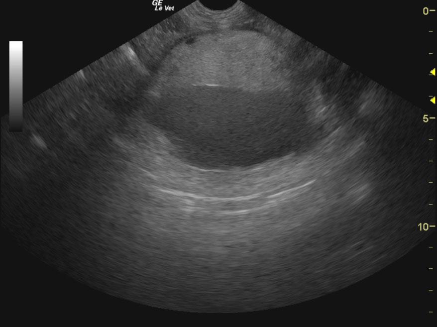
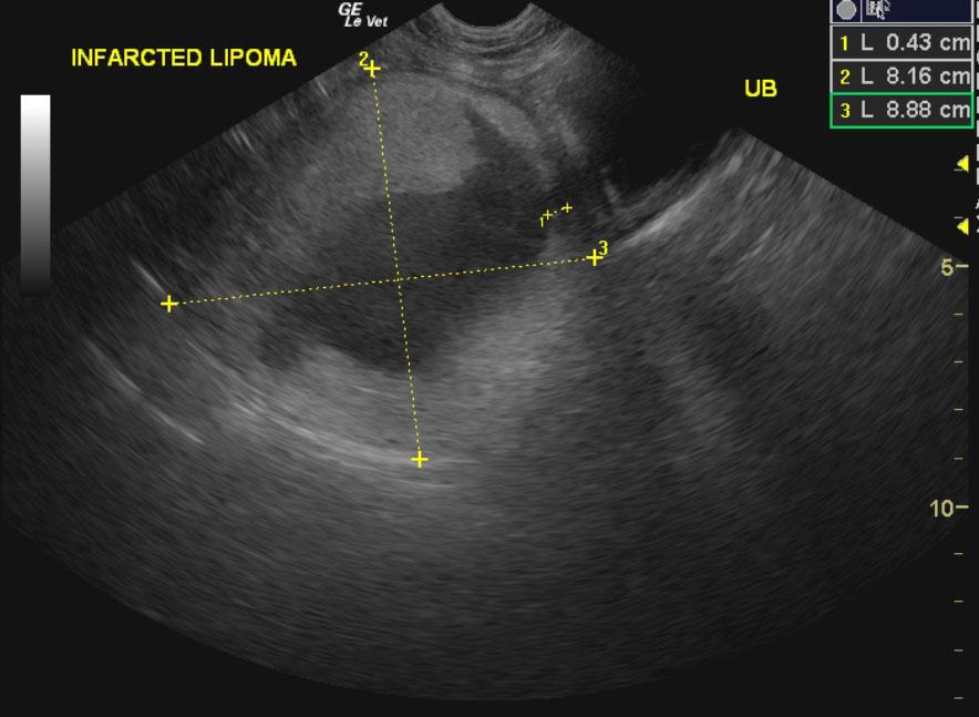
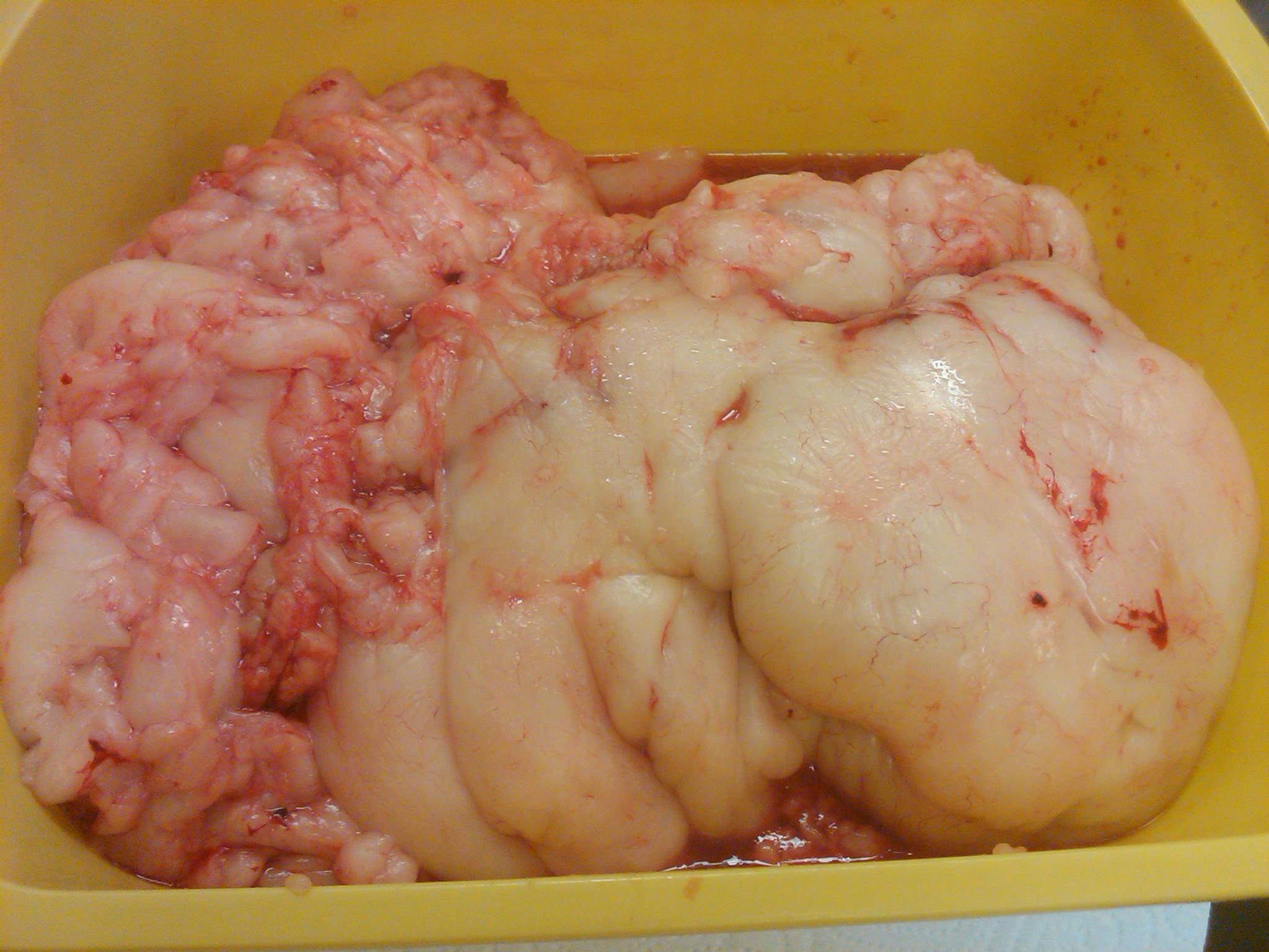
A 10-year-old FS Labrador Retriever dog was presented for boarding at rDVM. On physical examination, bilateral otitis, bilateral lenticular sclerosis, and a softball sized firm mass cranial to the bladder were present. Abdominal radiographs showed a spherical mass cranial to bladder and on the right side of abdomen. Abnormalities on urinalysis were 2+ proteinuria, 1+ bilirubinuria, few amorphous phosphates, and elevated microalbumin. CBC and blood chemistry were both within normal limits.
A 10-year-old FS Labrador Retriever dog was presented for boarding at rDVM. On physical examination, bilateral otitis, bilateral lenticular sclerosis, and a softball sized firm mass cranial to the bladder were present. Abdominal radiographs showed a spherical mass cranial to bladder and on the right side of abdomen. Abnormalities on urinalysis were 2+ proteinuria, 1+ bilirubinuria, few amorphous phosphates, and elevated microalbumin. CBC and blood chemistry were both within normal limits.