An 8-year-old MN domestic long hair cat was presented for vomiting, anorexia, depression, and diarrhea. Monocytosis, lymphopenia, and thrombocytopenia was evident on CBC, and serum biochemistry showed hypoalbuminemia, hypocalcemia, elevated AST activity, bilirubinemia, and hyperglycemia. T4 was within normal range. Possible sublumbar lymphadenomegaly and a small intestinal infiltrative disease leading to a partial obstruction was evident on survey radiographs.
An 8-year-old MN domestic long hair cat was presented for vomiting, anorexia, depression, and diarrhea. Monocytosis, lymphopenia, and thrombocytopenia was evident on CBC, and serum biochemistry showed hypoalbuminemia, hypocalcemia, elevated AST activity, bilirubinemia, and hyperglycemia. T4 was within normal range. Possible sublumbar lymphadenomegaly and a small intestinal infiltrative disease leading to a partial obstruction was evident on survey radiographs.
Case Study
Intestinal wall mass in a 8 year old MN DLH cat
Sonographic Differential Diagnosis
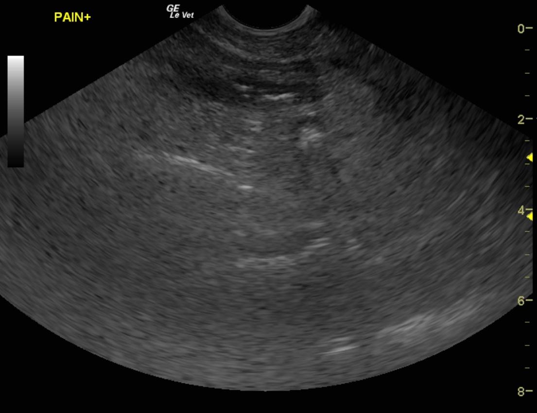
There is an intestinal wall mass with a potential for bowel infarction or complicating inflammatory bowel disease. IBD pattern elsewhere is evident with reactive omentum and mesenteric root lymph node. Necrotic intestine with potential for neoplasia is possible. Cholangiohepatitis and age-related renal changes was also evident.
Image Interpretation
The gastrointestinal tract presented prominent mucosa and a partially obstructive mid to distal small intestinal pattern with thickened, irregular hypoechoic wall with focal hyperechoic changes. The portion of thickened small intestine was causing a minor dilation with fluid. Underlying foreign body could not be visualized, but could not be ruled out either. This is consistent with gas penetration or potential mineralization. Mesenteric root lymphadenopathy was also noted. An adhesion pattern was noted associated with the other portions of small intestine. This is most consistent with complicated, inflammatory bowel disease or potential for emerging lymphoma or carcinoma. The pancreas presented mixed echogenic changes and appeared involved in the reactive omentum. A minor amount of free fluid and a significant amount of reactive omentum was noted.
DX
Outcome
The owner elected euthanasia.
Clinical Differential Diagnosis
Gastrointestinal disease (IBD, neoplasia, foreign body, granulomatous disease), pancreatic disease (pancreatitis, pancreatic neoplasia), liver disease (neoplasia, cholangiohepatitis complex, bacterial infection).
Sampling
None
Video
Patient Information
Clinical Signs
- Anorexia
- Depression
- Diarrhea
- Lethargy
- Vomiting
Images
Blood Chemistry
- Albumin, Low
- AST (SGOT), High
- Calcium, Low
- Glucose, High
- Total Bilirubin, High
CBC
- Lymphocytes, High
- Monocytes, High
- Platelet Count, Low
Clinical Signs
- Anorexia
- Depression
- Diarrhea
- Lethargy
- Vomiting