Intestinal necrosis and peritonitis with mineralized omental hematoma in a 7 year old M Labrador Retriever dog

Case Study

Intestinal necrosis and peritonitis with mineralized omental hematoma in a 7 year old M Labrador Retriever dog

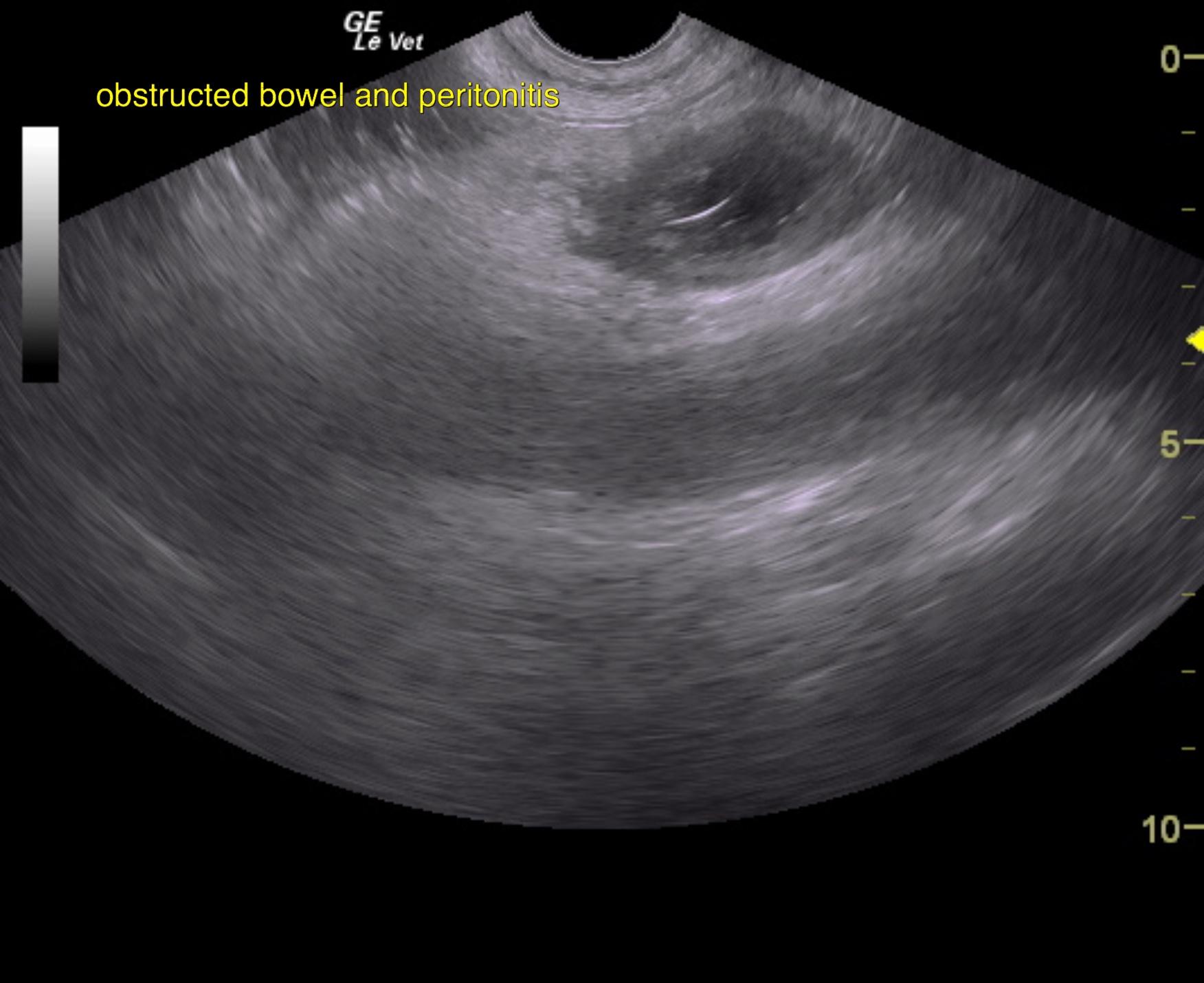
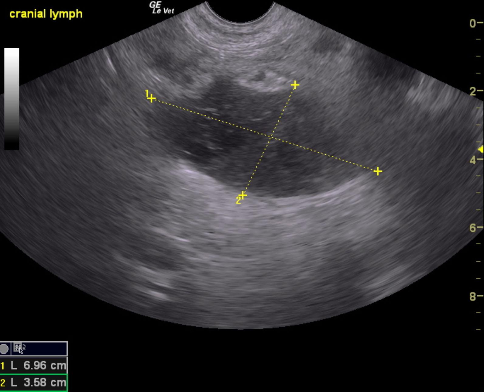
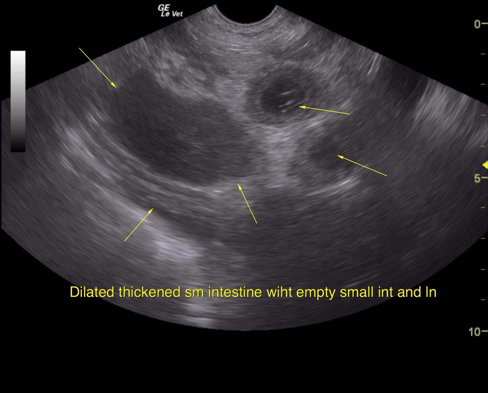
 A 7-year-old male Labrador with a history of previous foreign body ingestion was presented for evaluation of vomiting, anorexia, and lethargy. After 24 hours of symptomatic therapy the vomiting had stopped but there was ongoing anorexia. Radiographs showed a gas pattern but no obvious foreign body. Serosanginous fluid was aspirated on abdominocentesis.

Sonographic Differential Diagnosis

Distal small intestinal obstruction with unhealthy bowel and regional lymphadenopathy. Suspect underlying foreign body and perforation with regional peritonitis. Immediate exploratory surgery is recommended with expectations towards aggressive intestinal resection. Lymph node and intestinal wall sampling is essential as well as culture. Abdominal lavage will be necessary. Very guarded prognosis dependent upon underlying histopathology and outcome of surgery.

Image Interpretation

Variable portions of small intestine were thickened with loss of detail measuring 0.92 cm. Dilated intestine was noted followed by empty small intestine. A significant amount of regional mesenteric inflammation was noted. Some shadowing material was noted in either the colon or the distal small intestine; it was difficult to ascertain, as it was all coalesced together; however this creates an obstructive pattern. Mixed hypoechoic irregular lymph node was noted with hyperechoic surrounding fat and focal areas of cysts or necrosis.

DX

Surgical biopsies revealed intestinal necrosis and peritonitis with mild chronic lymphoplasmacytic enteritis and mineralized mental hematoma

Outcome

Surgery performed 8/21/15: torsion of the distal ileum to the cecum. Intestine black but no perforations noted. Hard mineralized mass removed from the momentum. Performed resection and anastomosis. Ravaged abdomen. Obtained sample of intestine and mineralized mass for biopsy. Released from the hospital on 8/23/15, returned for medical boarding until 8/31/15. Progress note 9/15/15, doing very well. Returned on 1/18/2016 for small laceration repair but otherwise doing very well since discharge.

Clinical Differential Diagnosis

Foreign body, focal perforation with peritonitis, necrotic enteritis, ulceration, neoplasia, intussusception

Sampling

Biopsy of the intestine revealed focal, severe transmural intestinal necrosis and peritonitis as well as mild chronic lymphoplasmacytic enteritis. Biopsy of the mass in the omentum revealed focal, nodular omental hematoma with mineralization.

Patient Information

Patient Name : Buddy Hammond/Animal Emergency/Dr. Fiflet/Andi
Gender : Male, Intact
Species : Canine
Type of Imaging : Ultrasound
Status : Complete
Liz Wuz Here : Yes
Code : 04_00505

Clinical Signs

  • Anorexia
  • Concern for FB Ingestion
  • Lethargy
  • Vomiting

Exam Finding

  • Dehydration
  • Underweight

Images

buddy_1buddy_2buddy_3

Blood Chemistry

  • ALT (SGPT), High

CBC

  • Neutrophils, High

Clinical Signs

  • Anorexia
  • Concern for FB Ingestion
  • Lethargy
  • Vomiting
Skip to content