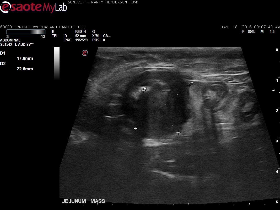
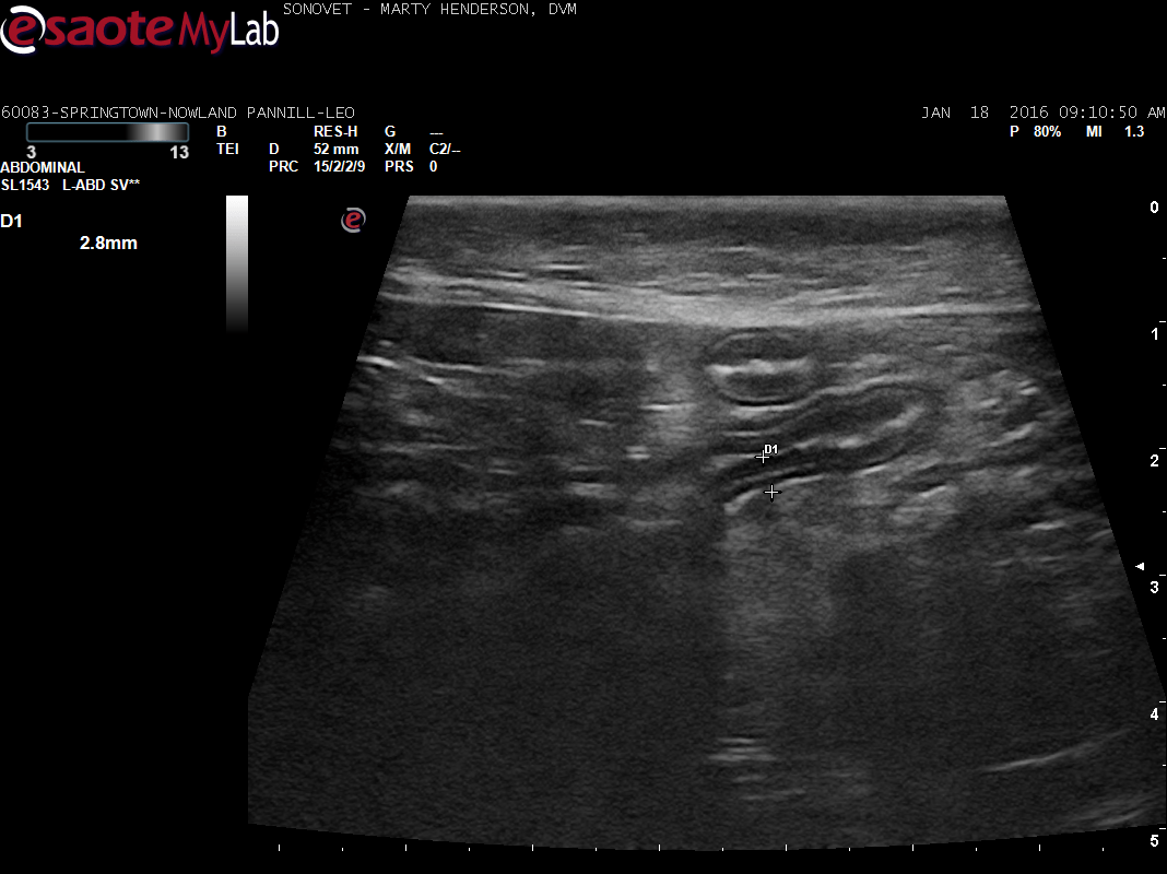
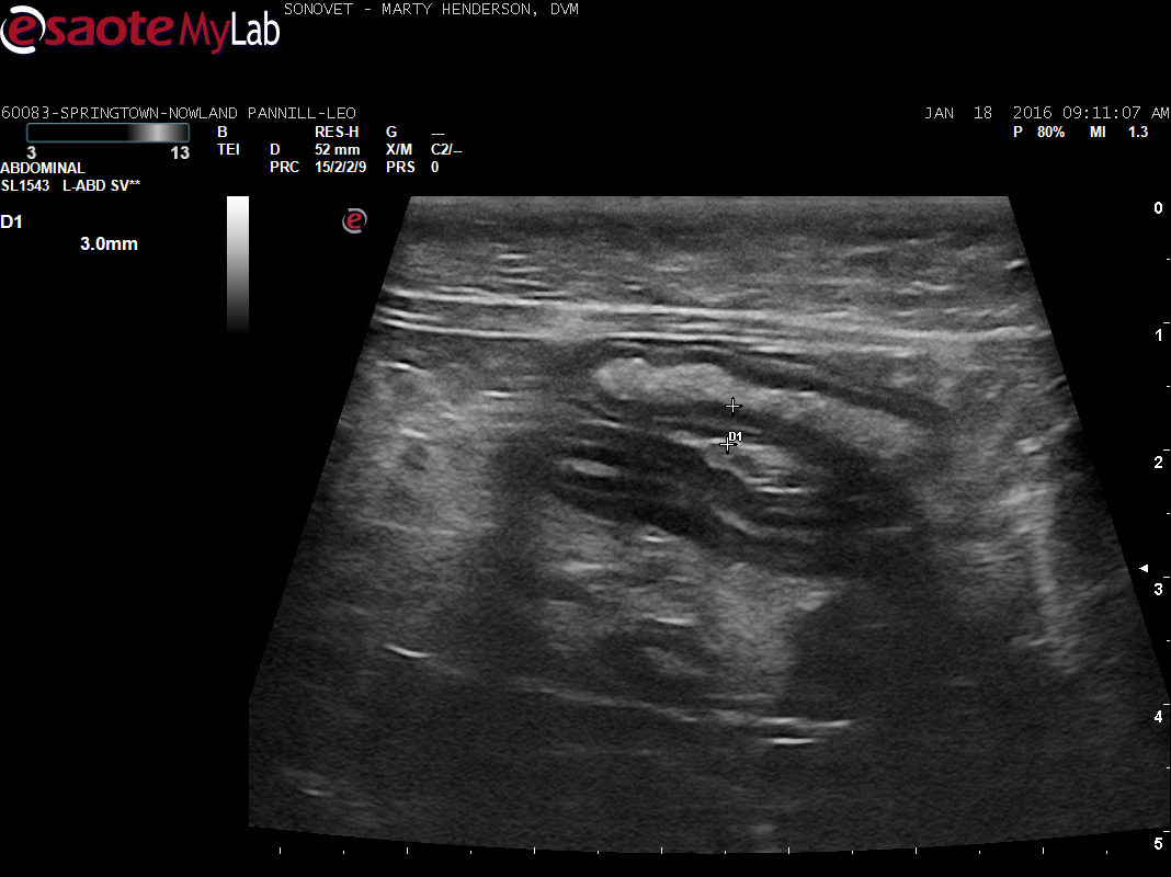
This 11 year old MN DSH cat vomited all food 3 days ago. Owner noticed grass-eating and some vomiting of grass last week. Normal activity and appetite. Currently on cerenia and SQ fluids.
R/O FB vs pancreatitis vs other
This 11 year old MN DSH cat vomited all food 3 days ago. Owner noticed grass-eating and some vomiting of grass last week. Normal activity and appetite. Currently on cerenia and SQ fluids.
R/O FB vs pancreatitis vs other