An 8-year-old FS Labrador Retriever dog with prior history of repeat gastrointestinal foreign bodies was presented for evaluation. The only significant abnormality on physical examination was a tense abdomen on palpation. CBC and serum chemistry were within normal limits. Survey abdominal radiographs demonstrated an abnormal gastrointestinal gas pattern.
An 8-year-old FS Labrador Retriever dog with prior history of repeat gastrointestinal foreign bodies was presented for evaluation. The only significant abnormality on physical examination was a tense abdomen on palpation. CBC and serum chemistry were within normal limits. Survey abdominal radiographs demonstrated an abnormal gastrointestinal gas pattern.
Case Study
Intestinal foreign body in an 8 year old FS Labrador Retriever dog
Sonographic Differential Diagnosis
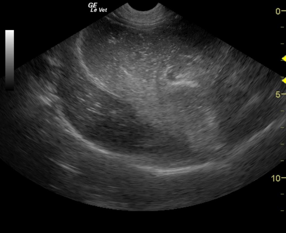
Obstructive small intestinal foreign body with likely linear gastric attachment.
Image Interpretation
The gastrointestinal tract presented a large small intestinal foreign body. It appeared to have a linear attachment into the stomach, potentially esophageal inlet. A large amount of dilation of the small intestine was noted prior to the cloth type foreign body. The small intestine was empty after the foreign body. A severe amount of gastric fluid was noted. A slight amount of free fluid was noted.
DX
Intestinal Foreign Body & IBD. Lymphoplasmacytic gastritis/enteritis.
Outcome
On exploratory laparotomy a foreign body was found in the ileum at the site of a previous anastomosis, which was removed via an enterotomy. Post surgery the dog was treated with Pepcid, polyflex, Cerenia, cefazolin, CRI of hetastarch and Lidocaine, and fed small amounts of Hills A/D. LRS. Abnormalities on CBC and serum biochemistry 48 hours post surgery were neutrophilia, monocytosis, and hypoalbuminemia. Two weeks post surgery the hypoalbuminemia had normalized and the owner reported that the dog was doing well at home.
Clinical Differential Diagnosis
GI tract pathology (foreign body, adhesions from previous foreign bodies/surgery, intestinal stenosis, IBD, neoplasia); Peritonitis; Pancreatic pathology (pancreatitis, neoplasia)
Sampling
Full thickness gastric and intestinal biopsies were taken. Mild, chronic, lympho-plasmacytic gastritis and moderate, chronic, lympho-plasmacytic and eosinophilic enteritis was diagnosed on histopathology.
Video
Patient Information
Patient Name :
Lucy G
Gender :
Female, Spayed
Species :
Canine
Type of Imaging : Ultrasound
Status :
Complete
Liz Wuz Here :
Yes
Code :
04_00240
Clinical Signs
- Concern for FB Ingestion
- Tense Abdomen
Exam Finding
- Tense Abdomen
Images
Clinical Signs
- Concern for FB Ingestion
- Tense Abdomen