This 16-year-old MN DSH cat presented for postprandial vomiting as well as diarrhea, weight loss, anorexia, and tenesmus. The physical examination revealed ropey intestines on abdominal palpation. A CBC and blood chemistry profile revealed a moderate neutrophilia with a left shift and normal chemistry values.
This 16-year-old MN DSH cat presented for postprandial vomiting as well as diarrhea, weight loss, anorexia, and tenesmus. The physical examination revealed ropey intestines on abdominal palpation. A CBC and blood chemistry profile revealed a moderate neutrophilia with a left shift and normal chemistry values.
Case Study
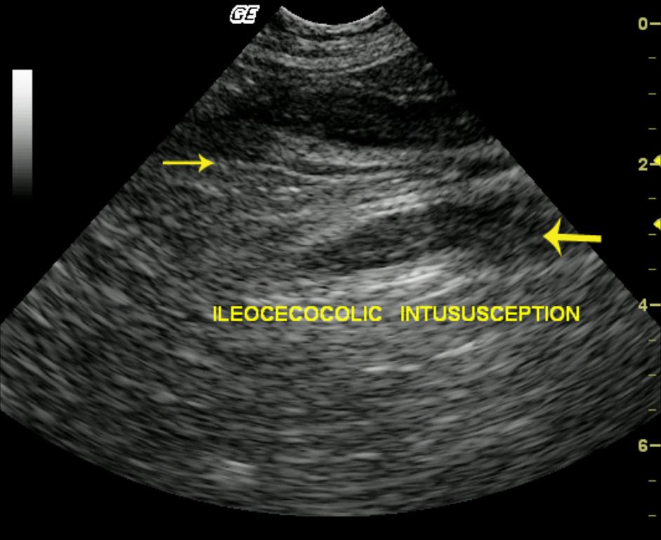
Ileocecocolic intussusception and chronic fibrosing enteritis in a 16 year old MN DSH cat
Sonographic Differential Diagnosis
Differentials for the primary disease included neoplasia and, less likely, inflammatory, or granulomatous disease.
Image Interpretation
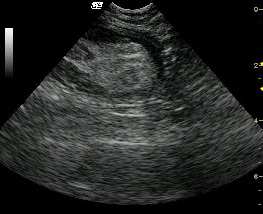
The imaged intestine showed a moderate to markedly, uniformly thick wall, which was hypoechoic with loss of wall layering (Videos 1 & 2.) The intestinal serosal layer was smooth and uninterrupted. The affected segment of bowel was mildly distended with echogenic material. A fusiform, hypoechoic focus with subtle layering was present in the center of the lesion. Video 3 demonstrates the location of the lesion at the level of the bladder trigone, continuing into the pelvic inlet; this is indicative of colonic involvement. An intussusception secondary to primary bowel wall disease was suspected.
DX
Ileocecocolic intussusception and chronic fibrosing enteritis
Outcome
The patient recovered uneventfully and was thriving 6 months post surgery.
Clinical Differential Diagnosis
Inflammatory bowel disease, neoplasia, concurrent pancreatitis, foreign body, FIP, intussusception, granulomatous disease.
Sampling
An exploratory laparotomy was performed, and a colectomy diagnosed the intussusception. Partial reduction with resection and anastomosis was done, as well as enteroplication. Histopathology revealed mild, chronic, fibrosing enteritis.
Video
Patient Information
Patient Name :
Tiger W
Gender :
Male, Neutered
Species :
Feline
Type of Imaging : Ultrasound
Book :
yes
Status :
Complete
Liz Wuz Here :
Yes
Code :
04_00047
Clinical Signs
- Diarrhea
- Tenesmus
- Vomiting
- Weight loss
Exam Finding
- Thickened Intestines
Images
CBC
- Left Shift
- Neutrophils, High
Clinical Signs
- Diarrhea
- Tenesmus
- Vomiting
- Weight loss