A 10-year-old Bernese Mountain Dog was presented for vomiting, anorexia, and diarrhea for 4 days. Physical examination was non-remarkable. Abnormalities on CBC and blood chemistry were leukocytosis, monocytosis, neutrophilia, lymphopenia, mild thrombocytopenia, hypoalbuminemia, and hypoproteinemia. Radiographs showed a prominent pylorus and some gas accumulation in the small intestines.
A 10-year-old Bernese Mountain Dog was presented for vomiting, anorexia, and diarrhea for 4 days. Physical examination was non-remarkable. Abnormalities on CBC and blood chemistry were leukocytosis, monocytosis, neutrophilia, lymphopenia, mild thrombocytopenia, hypoalbuminemia, and hypoproteinemia. Radiographs showed a prominent pylorus and some gas accumulation in the small intestines.
Case Study
IBD in a 10-year-old Bernese Mountain dog
Sonographic Differential Diagnosis
Minor gastrointestinal obstructive pattern possibly due to adhesion or dysfunctional bowel. Possible infiltrative disease such as early lymphoma.
Image Interpretation
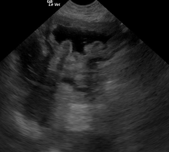
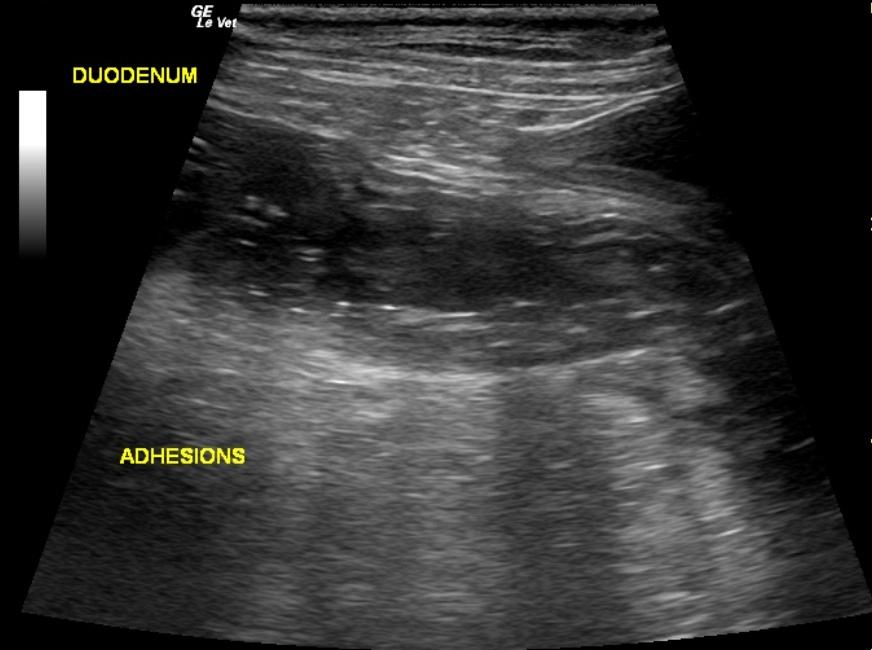
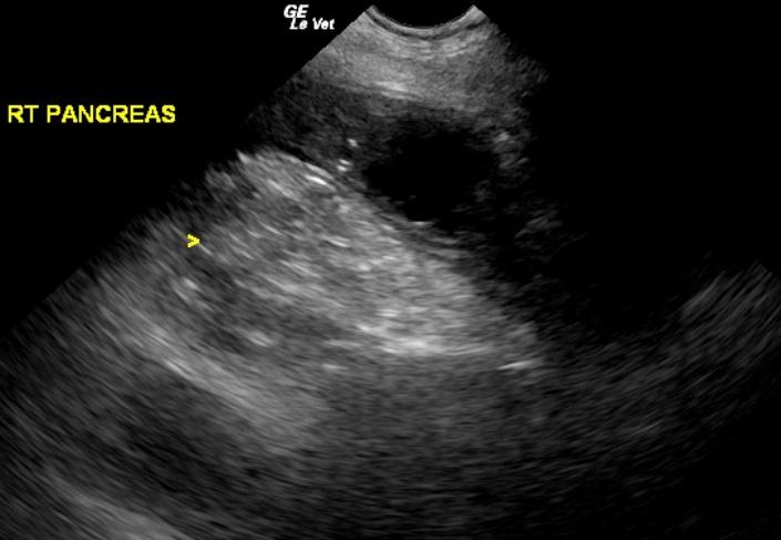
The stomach in this patient was dilated with edematous wall that continued into the duodenum. Anechoic luminal fluid was present. The descending duodenum was dilated yet peristaltic with multiple areas of adhesions continuing out of the area of the pancreas. Some mixed hypoechoic changes consistent with pancreatitis were also present, however this is considered part of the process and not the primary cause. Some omental inflammation continued into the area of the right kidney which was also swollen and painful. The small intestinal dilation continued into the mid jejunum which was empty at that point and aperistaltic. This is an obstructive pattern, however no overt foreign body could be seen other than some artifact. Transmural pathology is suspected given that some omental adhesions were present along the affected small intestines with gas penetration into the wall.
DX
Outcome
The patient was treated post-operatively with I.V. fluids, famotidine, baytril, and buprenex and discharged several days later with baytril, metronidazole, pepcid, rimadyl, and eventual prednisone. At suture removal the patient was doing well on the current medication.
Comments
Full thickness biopsies as well as liberation of any dysfunctional bowel or potential bowel infarction is highly recommended.
Clinical Differential Diagnosis
GI tract pathology (neoplasia, foreign body, IBD, motility disorder, ulceration). Pancreatic pathology (pancreatitis, neoplasia). Peritonitis. Liver pathology (chronic liver disease, neoplasia).
Sampling
Full-thickness surgical biopsies of the ascending duodenum and the distal tip of the right side of the pancreas were performed. Biopsy of the pancreas revealed an islet tumor whereas the duodenum revealed moderate lymphoplasmacytic-eosinophilic enteritis, suggestive of IBD.
Video
Patient Information
Clinical Signs
- Anorexia
- Diarrhea
- Vomiting
- Weight loss
Images
Blood Chemistry
- Albumin, Low
- Total Protein, Low
CBC
- Lymphocytes, Low
- Monocytes, High
- Neutrophils, High
- Platelet Count, Low
- WBC, High
Clinical Signs
- Anorexia
- Diarrhea
- Vomiting
- Weight loss