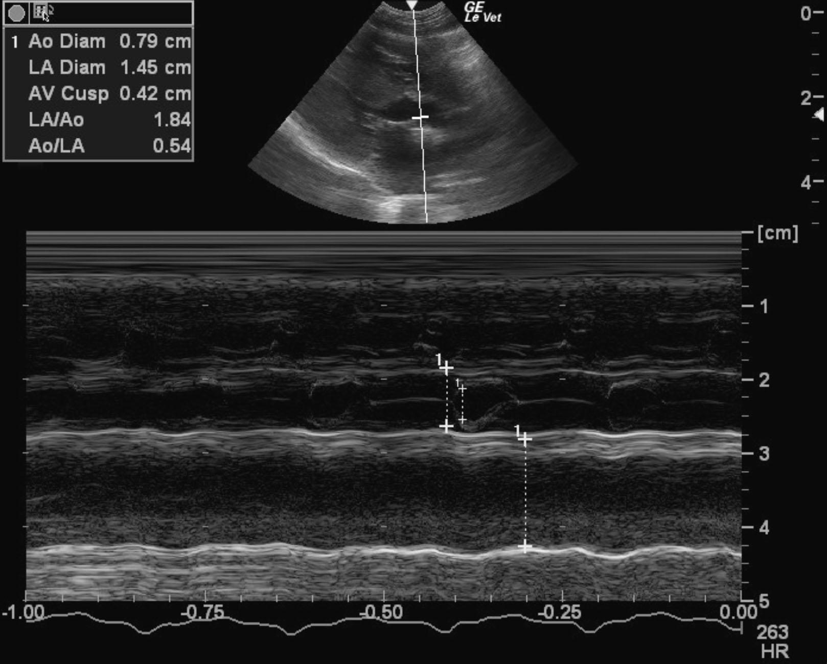
A 1-year-old MN DSHcat with a history of cardiac disease that was being treated with Plavix, enalapril, pimobendan, furosemide, and atenolol was presented for evaluation of weakness and dyspnea.
Physical exam: tachycardia (200-400bpm) and a grade III/VI heart murmur
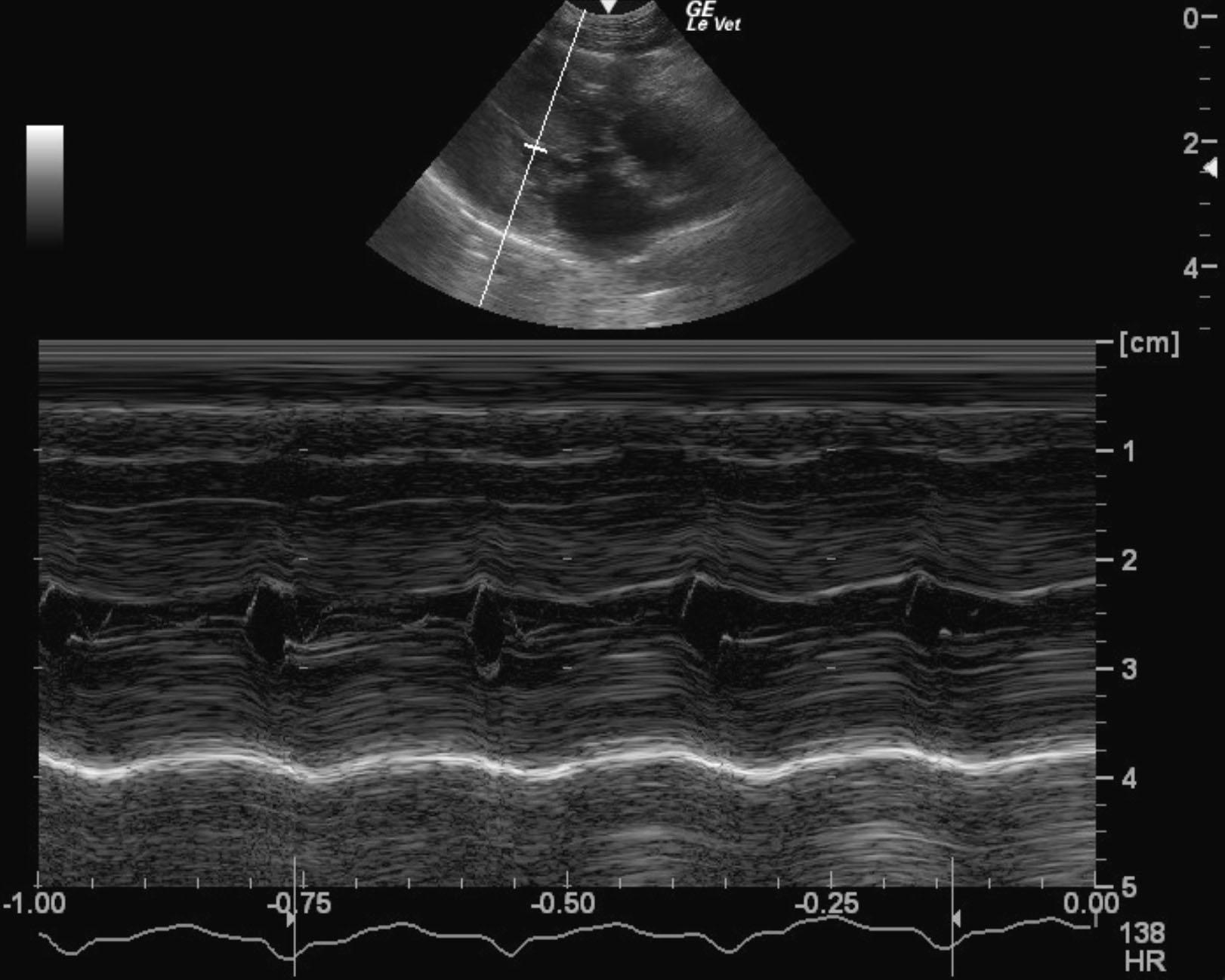
A 1-year-old MN DSHcat with a history of cardiac disease that was being treated with Plavix, enalapril, pimobendan, furosemide, and atenolol was presented for evaluation of weakness and dyspnea.
Physical exam: tachycardia (200-400bpm) and a grade III/VI heart murmur