Hyperechoic mucosal layer in a 6-year-old FS Havanese: A forum post.

Case Study

Hyperechoic mucosal layer in a 6-year-old FS Havanese: A forum post.

A 6-year FS Havanese with history of vomitng (mostly at night and in morning) for a few weeks; no diarrhea. Recent bloodwork found ALB 16, TP 40, GLOB 24 with other parmaters WNL. A fecal was negative. Urine SG 1.035, no protein on chemstrip and urine pro:crea ratio was pending.

A 6-year FS Havanese with history of vomitng (mostly at night and in morning) for a few weeks; no diarrhea. Recent bloodwork found ALB 16, TP 40, GLOB 24 with other parmaters WNL. A fecal was negative. Urine SG 1.035, no protein on chemstrip and urine pro:crea ratio was pending.

Image Interpretation

Ultrasound showed an approx. 4cm length of small intestine with a uniformly hyperechoic mucosal layer with overall normal wall thickness and normal layering; the classic mucosal striations were not seen, but some mucosal speckling was present.

DX

Severe primary Lymphangiectasia.

Outcome

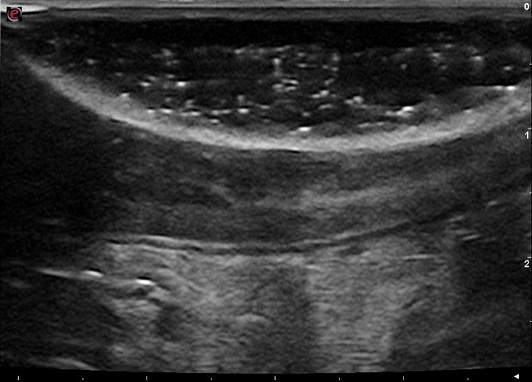
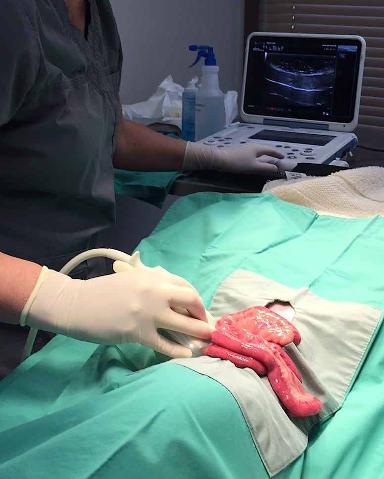
“At first I thought it was my probe or settings but then I switched presets and also compared other loops of bowel which had a normal hypoechoic mucosal layer so I do believe this lesion is real – rest of scan was normal; no effusion or enlarged LN’s. So looks like a protein losing enteropathy (pending normal pro:crea ratio). What are your thoughts of this bowel lesion? I am scheduled to perform intra-operative ultrasound on this as I think it may be difficult to determine where it is grossly. The plan is to identify the abnormal loop of bowel and biopsy or possibly resect.” Histopath Results: These intestinal biopsies are excellent, and are definitive for primary lymphangiectasia.
The intestine is structurally normal and has no increase in leukocytes, but every villus lacteal is
massively distended to the point of rupture, and the dilation of lymphatics continues within the
submucosa and tunica muscularis. There is absolutely no evidence of neoplasia, and there is no
evidence of previous inflammation or fibrosis that might explain the development of the
lymphangiectasia. The vast majority of cases of lymphangiectasia that I see in dogs are primary
and idiopathic like this.

Comments

This case study was sampled from SonoPath’s interactive forum, to see the full forum thread (it’s a good one!) click here. 🙂

Sampling

Full-thickness intestinal biopsies were sampled via intraoperative ultrasound.

UA Specific Gravity Range

1.035

Patient Information

Gender : Female, Spayed
Species : Canine
Type of Imaging : Ultrasound
Status : Complete

Clinical Signs

  • "Not Doing Right"
  • Diarrhea
  • Vomiting

Images

small_intestine_specklinglympahgiectsasiasi_intraop_img_2090

Clinical Signs

  • "Not Doing Right"
  • Diarrhea
  • Vomiting
Skip to content