A 13-year-old MN Welsh Corgi dog was presented for the evaluation of a decreased appetite and to check his left hind leg. The dog had a history of discoid lupus and alopecia on the chin. The owner had also reported that the dog was having urinary accidents in the house. The dog had an elevated temperature on physical examination, as well as bilateral cataracts. His hind limbs were splayed and he was painful upon palpation of the mid-lumbar spine and the left carpus.
A 13-year-old MN Welsh Corgi dog was presented for the evaluation of a decreased appetite and to check his left hind leg. The dog had a history of discoid lupus and alopecia on the chin. The owner had also reported that the dog was having urinary accidents in the house. The dog had an elevated temperature on physical examination, as well as bilateral cataracts. His hind limbs were splayed and he was painful upon palpation of the mid-lumbar spine and the left carpus. Blood chemistry revealed an increased urea, hypoalbuminemia, hypercholesterolemia, hyperamylasemia, an elevated GGT enzyme activity and hyperphosphatemia. Lymphopenia was present on the CBC. Urinalysis showed proteinuria (2+), hematuria (+1), high RBC (11-20/HPF), and the presence of bacteria (cocci and rods 2+). The urine microalbumin was strongly positive. The urine culture was negative. Lyme C6В® quantitative test result was negative.
Case Study
Hepatitis with focal nodular hyperplasia in a 13 year old MN Welsh Corgi dog
Sonographic Differential Diagnosis
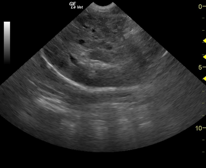
Undefined deep left medial liver mass. Hepatocellular carcinoma or other neoplasia likely. Chronic lobar necrosis or granulomatous lesion possible.
Image Interpretation
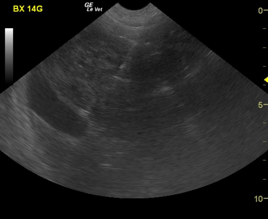
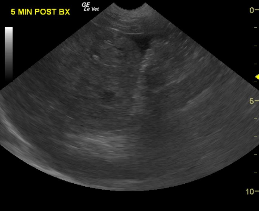
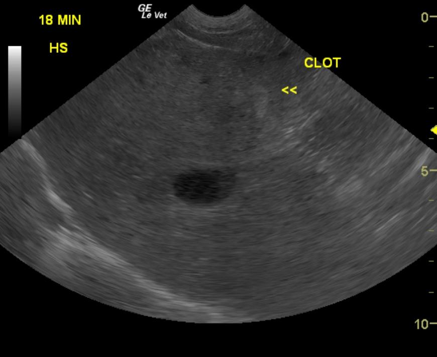
The liver presented some echogenic remodeling in both left and right lobes, however a 6cm highly vascular mass was noted in the deep left lobe adjacent to the gallbladder. The common bile duct and portal vein were not overtly affected however impingement upon the gallbladder and cystic duct was noted. Resection may be somewhat tedious with full left liver lobectomy. US-guided core biopsy (14-gauge) was performed of the liver mass with evident sampling hemorrhage indicated by the anechoic free fluid in the near field in images 3. This stabilized after 20-30 minutes as a clot formed (image 4)
DX
Outcome
Coagulation panel was within normal limits and a recheck CBC showed mild anemia; the PCV/TP were 28%/5.2 g/dL, respectively. Patient underwent a lobectomy of the liver. The first sample taken from the liver mass revealed nodular hyperplasia with hematoma formation, infarction and moderate chronic portal hepatitis with moderate bridging fibrosis. The second liver biopsy showed moderate chronic hepatitis with focal nodular hyperplasia. The patient was pyrexic a few hours postoperatively, but normalized by the next morning. He remained hospitalized on I.V. fluids and supportive care. The patient’s appetite gradually returned and he was discharged with oral antibiotics, hepatoprotectants, steroids, and a gastroprotectant. At recheck exam, he was eating well and had pink mucous membranes.
Clinical Differential Diagnosis
Urinary accidents possibly secondary to polydipsia and polyuria (with the polydipsia not having been noted by the owner); renal insufficiency, pyelonephritis, GN, or neoplasia should be considered. In addition, hepatic disease, such as chronic active hepatitis, cholestasis, cholangitis, infectious hepatopathy (leptospirosis) or neoplasia must be considered. Examples include a hepatocellular adenoma, carcinoma, lymphoma, leiomyoma, leiomyosarcoma, histiocytic sarcoma, mast cell tumor, etc. The back and left carpal pain could be due to intervertebral disk disease, or osteoarthritis, respectively. Other diseases such as polyarthritis (back pain caused by inflammation of the articular facets) associated with a possible recurrence of the systemic lupus erythematosus may be considered. Other causes of back pain include discospondylitis (fever.)
Sampling
US-guided Tru-cutВ® biopsy and full-thickness surgical biopsies. US-guided core biopsy: Taken from the liver mass revealed nodular hyperplasia with hematoma formation, infarction and moderate chronic portal hepatitis with moderate bridging fibrosis. Surgical biopsy: Moderate chronic hepatitis with focal nodular hyperplasia.
Video
Patient Information
Clinical Signs
- Anorexia
- Inappropriate Urination
Exam Finding
- Cataracts
- Fever
- Pain
Images
Blood Chemistry
- Albumin, Low
- Amylase, High
- BUN high
- Cholesterol, High
- GGT High
- Phosphorus, High
CBC
- Lymphocytes, Low
- WBC, Low
Clinical Signs
- Anorexia
- Inappropriate Urination
Urinalysi
- Albumin Present
- Bacteria Present
- Blood Present
- Culture negative
- Protein Present