Hemorrhagic lung mass found in transdiaphragmatic view while assessing for pancreatitis in 9 year old MN Bassett Hound

Case Study

Hemorrhagic lung mass found in transdiaphragmatic view while assessing for pancreatitis in 9 year old MN Bassett Hound

A 9-year-old MN Bassett Hound was presented for history of possible pancreatitis, lethargy, and anorexia, which was progressive over the last week. Abnormalities on CBC and blood chemistry were marked leukocytosis, left shift neutrophilia, lymphopenia, mild monocytosis, eosinopenia, and hyperamylasemia. Spec cPL was elevated. Abdominal radiographs showed possible ileus with no foreign body or masses evident. The patient was treated with IV fluids, antibiotics, and analgesics.
A 9-year-old MN Bassett Hound was presented for history of possible pancreatitis, lethargy, and anorexia, which was progressive over the last week. Abnormalities on CBC and blood chemistry were marked leukocytosis, left shift neutrophilia, lymphopenia, mild monocytosis, eosinopenia, and hyperamylasemia. Spec cPL was elevated. Abdominal radiographs showed possible ileus with no foreign body or masses evident. The patient was treated with IV fluids, antibiotics, and analgesics.
Sonographic Differential Diagnosis

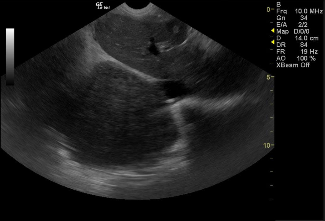
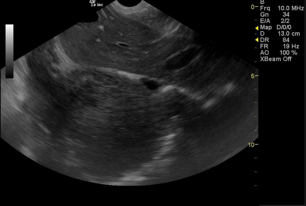
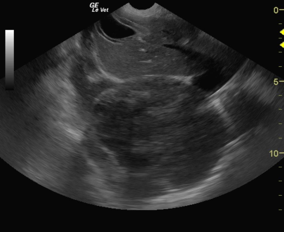
These findings were most consistent with dorsal caudal lung sarcoma with potential of lung lobe torsion. Necrosis and hemorrhage associated with the mass makes this a surgical urgency. However, full resection is unlikely to be possible. Computed tomography can further define this change.

Image Interpretation

While examining the cranial abdomen and caudal thorax (transdiaphragmatic), the sonographer noted a minor amount of free pleural fluid and a large mixed echoic thoracic mass. Further investigation with thoracic ultrasound revealed an 8cm abscessing caudal lung mass, which is likely lung sarcoma.

DX

Suggestive for lung neoplasia

Outcome

The owner elected euthanasia.

Comments

It is essential for the sonographer to complete a full abdominal study and image the caudal thorax through the diaphragm. This manuever changed the direction completely in this case that revealed minimal abdominal pathology.

Clinical Differential Diagnosis

Acute pancreatitis, pancreatic abscessation, pancreatic neoplasia, and focal peritonitis secondary to intestinal perforation.

Sampling

An US-guided FNA of the mass in the lungs revealed ghost cells with necrosis, most likely neoplastic. The free fluid was also sampled and found to be hemorrhagic.

Patient Information

Patient Name : Toby P
Gender : Male, Neutered
Species : Canine
Type of Imaging : Ultrasound
Book : yes
Status : Complete
Liz Wuz Here : Yes
Code : 10_00025

Clinical Signs

  • Anorexia
  • Lethargy

Images

PetersonTobyk9lungmassthoracic_01132012080212PetersonTobyk9lungmassthoracic2_01132012080225PetersonTobyk9lungmassthoracic3_01132012080238

Blood Chemistry

  • Amylase, High

CBC

  • Eosinophils, High
  • Left Shift
  • Lymphocytes, Low
  • Monocytes, High
  • WBC, High

Clinical Signs

  • Anorexia
  • Lethargy

Special Testing

  • cPLI Positive