Granulosa cell ovarian tumor (diagnosed by FNA) and incidental right adrenal tumor in a 13 year old FI Fox Terrier

Case Study

Granulosa cell ovarian tumor (diagnosed by FNA) and incidental right adrenal tumor in a 13 year old FI Fox Terrier

A 13 year old intact female Fox terrier was presented for evaluation of possible pyometra or abdominal mass. A mid abdominal thickening was palpable.
A 13 year old intact female Fox terrier was presented for evaluation of possible pyometra or abdominal mass. A mid abdominal thickening was palpable.
Sonographic Differential Diagnosis

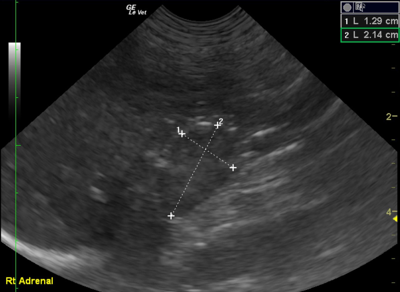
The left ovarian tumor appears resectable. The right adrenal gland mass is potentially resectable. Differentials for the right adrenal mass include adenocarcinoma or pheochromocytoma, given the invasive activity. Adrenal adenoma with attached thrombosis is also possible.

Image Interpretation

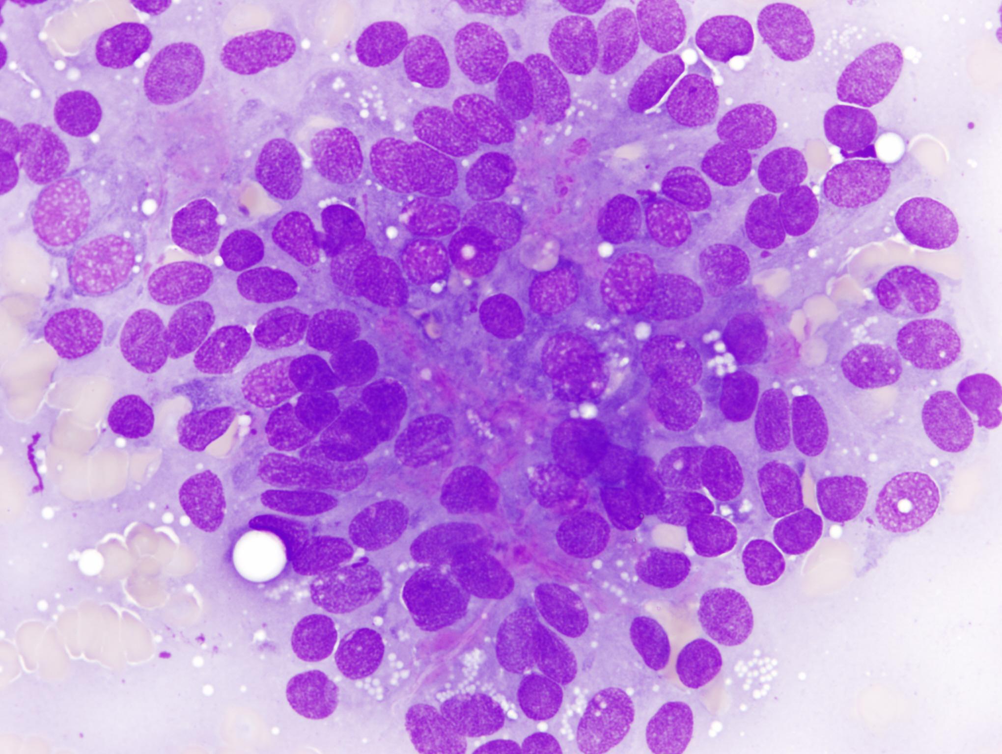
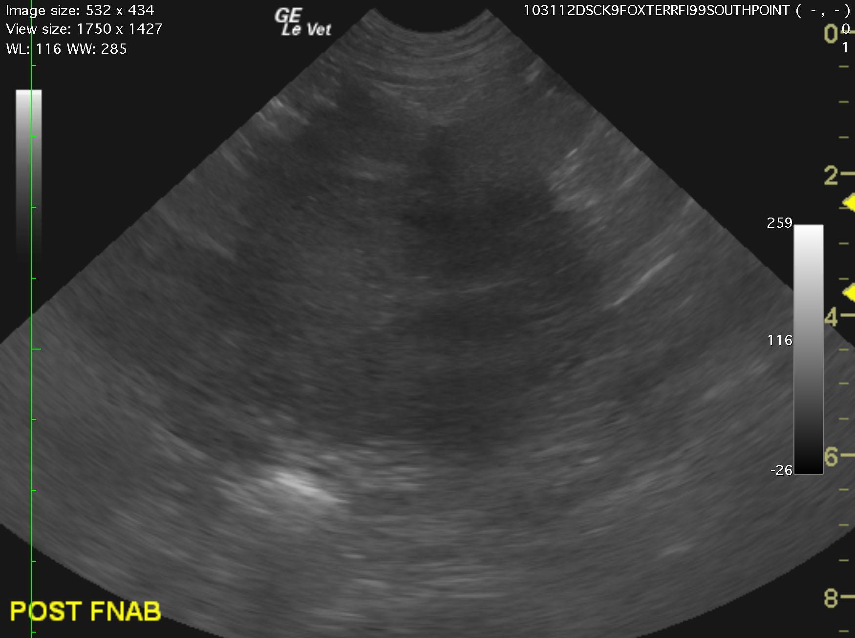
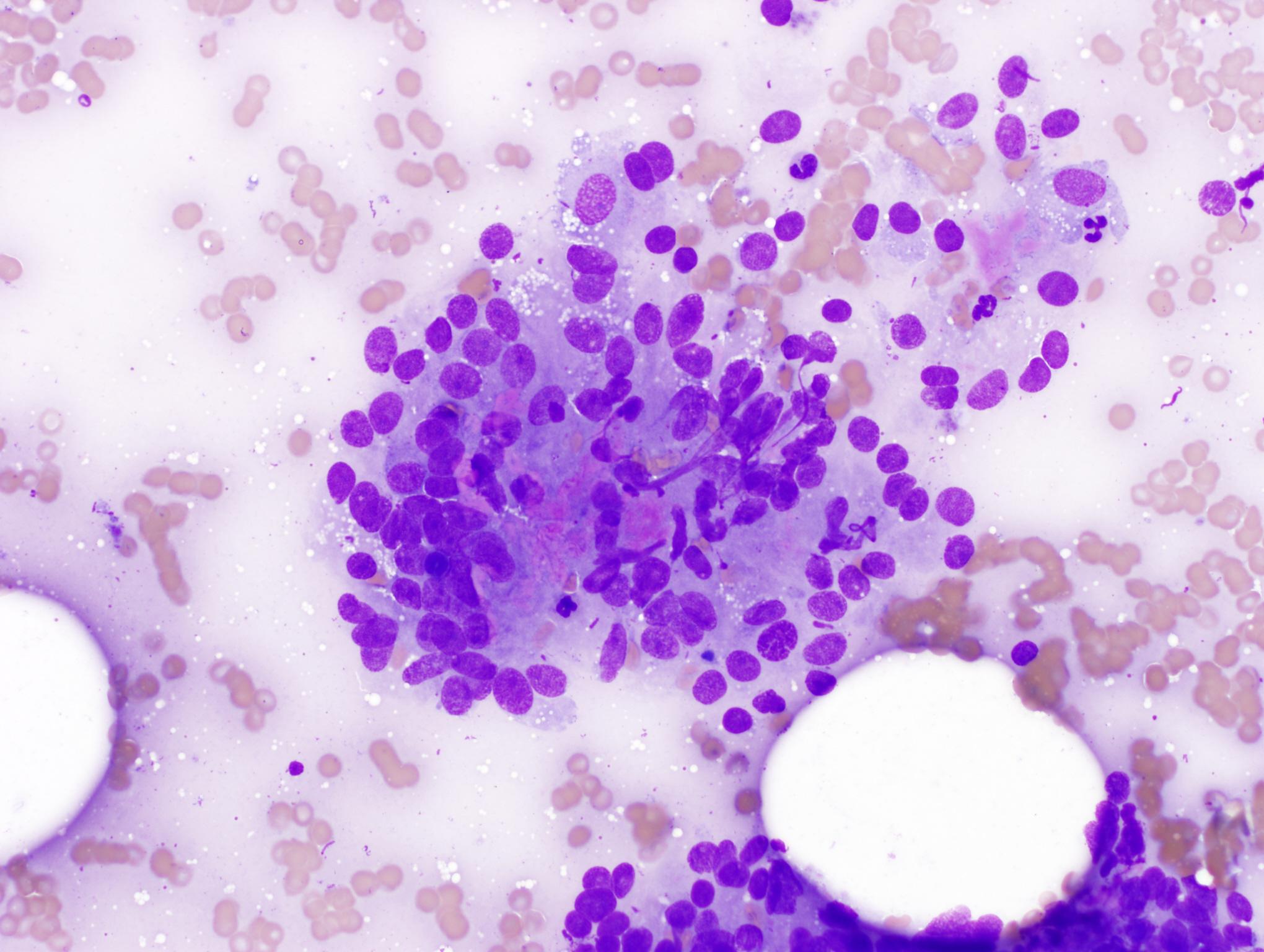
The left ovary presented a cystic portion that measured 2.0 cm with an attached mass that measured 5.57 x 4.92 cm (Image 1 and videos 1 and 2). This is consistent with a left ovarian tumor. Carcinoma or granulosa cell tumor is suspected. The region of the right ovary was unremarkable. The uterine body was unremarkable. The left adrenal gland was uniform and measured 0.58 cm (not shown). The right adrenal gland was mildly enlarged and slightly irregular which may be normal for this patient given the age. The right adrenal measured 2.14 x 1.29 cm. After further imaging of the CVC and right phrenic vein, the irregular right adrenal gland appeared to slightly invade the vena cava with attached caval thrombus or caval extension of the tumor (Video 2). Images 3 and 4 reveal 40x and 100x cytology of the left ovarian granulosa cell tumor obtained by 25 gauge US-guided FNA.

DX

Granulosa cell tumor of the ovary. Unilateral adrenomegaly.

Outcome

Recommend ovariohysterectomy and right adrenalectomy in this patient. CT evaluation of the right adrenal could also be considered. Blood pressure measurements and full adrenal gland panel is recommended. The owners are considering their options.

Comments

Why do we always perform a full abdominal sonogram from diaphragm to pelvic urethra? Simple ovarian tumor and eventual OVH? Even though it may seem so, the prudent clinical sonographer will image the right adrenal gland in full (even if not overtly indicated from the history) and not be satisfied with the simple 2-D right adrenal view. Dr. Doug Casey of English Bay Ultrasound Service, Vancouver, B.C, Canada (http://www.sonopath.com/specialists_casey.asp) gives the practitioner and client something more to think about in this month’s SonoPath November 2012 case of the month.

Clinical Differential Diagnosis

Mass – neoplasia, granuloma, abscess or cyst (in any of the following organs: spleen, kidney, liver, ovary, intestine, mesentery, or lymph node). Hydronephrosis. Splenic torsion. Pyometra.

Sampling

US-guided FNA of the parenchymal portion of the ovarian mass was performed without complication. Cytology revealed granulosa cell tumor of the ovary.

Patient Information

Patient Name : Pepsi T
Gender : Female, Intact
Species : Canine
Type of Imaging : Ultrasound
Status : Complete
Liz Wuz Here : Yes
Code : 09_00052

Clinical Signs

  • Palpable Mass

Exam Finding

  • Palpable mass

Images

pepsi1copy_11032012114208pepsi4_11032012114605pepsi7_11032012114723pepsi8_11032012114746

Clinical Signs

  • Palpable Mass