This 9-year-old FS Old English Sheepdog was presented for persistent vomiting and anorexia. Physical exam of the dog revealed normal CBC and blood chemistry analysis.
This 9-year-old FS Old English Sheepdog was presented for persistent vomiting and anorexia. Physical exam of the dog revealed normal CBC and blood chemistry analysis.
Case Study
Gastric Foreign Body in a 9 year old Old English Sheepdog
Sonographic Differential Diagnosis
Uncomplicated gastric foreign body.
Image Interpretation
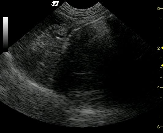
The imaged segment of stomach is markedly distended with an ovoid, 3 cm structure possessing fragmented distal acoustic shadowing. In the near field, normal bowel wall layering is preserved and adjacent peritoneal inflammation or free fluid is not visualized suggesting a healthy segment of stomach.
DX
Gastric foreign body
Outcome
Exploratory gastrotomy revealed a conglomeration of plastic ball casing, rubber tubing, food and hair.
Clinical Differential Diagnosis
GI pathology – Gastroenteritis, IBD, neoplasia, GI foreign body, GI ulceration; Pancreatic pathology – pancreatitis; atypical Addison’s disease, toxicosis, infectious.
Video
Patient Information
Patient Name :
Bella W
Gender :
Female, Spayed
Species :
Canine
Type of Imaging : Ultrasound
Status :
Complete
Liz Wuz Here :
Yes
Code :
04_00097
Clinical Signs
- Anorexia
- Vomiting
Images
Clinical Signs
- Anorexia
- Vomiting