An 8-year-old MN DSH cat with a history of a palpable abdominal mass and anorexia, was presented for evaluation. CBC was within normal limits whereas on serum chemistry hypercholesterolemia (414) and hyperamylasemia (1327) was present. Survey abdominal radiographs showed a large opacity in the cranial abdomen.
An 8-year-old MN DSH cat with a history of a palpable abdominal mass and anorexia, was presented for evaluation. CBC was within normal limits whereas on serum chemistry hypercholesterolemia (414) and hyperamylasemia (1327) was present. Survey abdominal radiographs showed a large opacity in the cranial abdomen.
Case Study
Gastric foreign body in an 8 year old MN DSH cat
Sonographic Differential Diagnosis
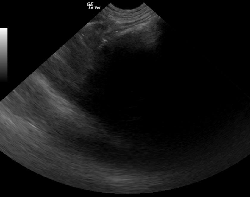
Large gastric foreign body, trichobezoar suspected but other foreign material possible.
Image Interpretation
Intraluminal gastric hyperechoic near field border with distal acoustic shadowing. Note some variability in near field sound absorption to the right side of the image.
DX
Gastric foreign body
Outcome
A gastrotomy was performed and a large amount of ribbon and shoelace removed. Post surgery recovery was uneventful.
Comments
No video is available on this patient.
Clinical Differential Diagnosis
Abdominal mass – Liver pathology (neoplasia, cyst, granuloma); Renal pathology (neoplasia, hydronephrosis); GI pathology (foreign body, neoplasia); Mesenteric pathology (neoplasia, granuloma); Pancreatic pathology (neoplasia, granuloma)
Sampling
None
Patient Information
Patient Name :
Ripken F
Gender :
Male, Neutered
Species :
Feline
Type of Imaging : Ultrasound
Status :
Complete
Liz Wuz Here :
Yes
Code :
04_00235
Clinical Signs
- Anorexia
Exam Finding
- Palpable mass
Images
Blood Chemistry
- Amylase, High
- Cholesterol, High
Clinical Signs
- Anorexia