This 12-year-old FS Portuguese Water Dog presented for acute onset of anorexia, lethargy, vomiting and diarrhea. The physical exam revealed hematochezia present on rectal exam with mild dehydration and distended stomach.
This 12-year-old FS Portuguese Water Dog presented for acute onset of anorexia, lethargy, vomiting and diarrhea. The physical exam revealed hematochezia present on rectal exam with mild dehydration and distended stomach.
Case Study
Gastric Foreign Body in a 12 year old FS Portuguese Water Dog
Sonographic Differential Diagnosis
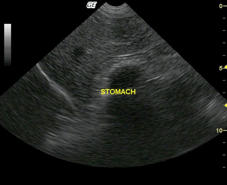
Currently non-obstructing gastric foreign body.
Image Interpretation
An ovoid, significantly shadowing gastric luminal foreign body is present. The foreign body interface is fragmented and indistinct supporting a rough, irregular surface. The foreign body appears to be within the gastric fundus and non-obstructing at the time of ultrasound. The remaining gastric lumen is moderately filled with echogenic fluid and ingesta. Based on the history intermittent pyloric outflow obstruction is likely occurring.
DX
Gastric foreign body
Outcome
The patient recovered uneventfully and was doing well at a 1-month follow-up examination
Clinical Differential Diagnosis
GI pathology – Gastroenteritis, IBD/colitis, infectious, bowel infarction, HE, foreign body, neoplasia.
Sampling
Gastrotomy revealed a peach pit located in the gastric lumen
Video
Patient Information
Patient Name :
Zelda R
Gender :
Female, Spayed
Species :
Canine
Type of Imaging : Ultrasound
Status :
Complete
Liz Wuz Here :
Yes
Code :
04_00065
Clinical Signs
- Anorexia
- Diarrhea
- Lethargy
- Vomiting
Exam Finding
- Abdominal Distension
- Dehydration
- Fresh Blood in Stool
Images

Clinical Signs
- Anorexia
- Diarrhea
- Lethargy
- Vomiting