An 11-year-old mixed breed dog was presented with a 2 week history of vomiting but with a good appetite. The only abnormality on physical examination was a tense abdomen on palpation. Rectal exam revealed gas and a small amount of stool. Fecal examination and a Giardia Elisa Snap test were both negative. Abnormalities on CBC and serum biochemistry were mild thrombocytosis, elevated ALP and elevated GGT activity, hyperkalemia, and mild hypocalcemia. On survey abdominal radiographs a poorly defined 2cm oblong soft tissue density was evident.
An 11-year-old mixed breed dog was presented with a 2 week history of vomiting but with a good appetite. The only abnormality on physical examination was a tense abdomen on palpation. Rectal exam revealed gas and a small amount of stool. Fecal examination and a Giardia Elisa Snap test were both negative. Abnormalities on CBC and serum biochemistry were mild thrombocytosis, elevated ALP and elevated GGT activity, hyperkalemia, and mild hypocalcemia. On survey abdominal radiographs a poorly defined 2cm oblong soft tissue density was evident.
Case Study
Gastric foreign body in an 11 year old mixed breed dog
Sonographic Differential Diagnosis
Stomach: Inflammatory or neoplastic infiltrative mucosal change. Possible foreign material or focal inflammation with surface ulceration. Pancreatic body and right limb- inflammatory or neoplastic infiltrative change, possibly early amyloid infiltrate (could be normal for this patient).
Image Interpretation
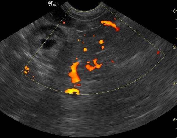
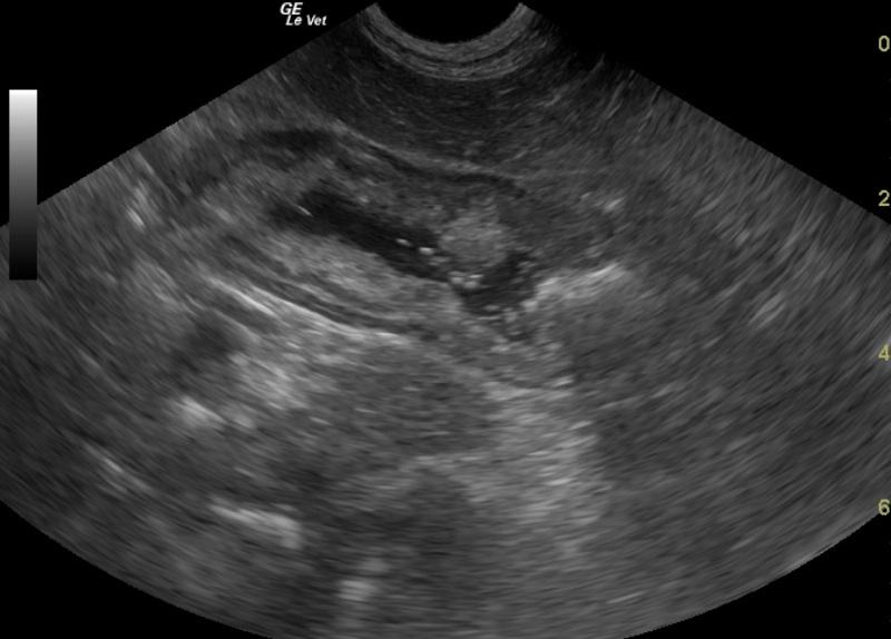
Thickened, prominent, mildly irregular pancreatic body and thickened stomach. Vascularity of pancreas imaged using Doppler. Thickened stomach wall, greater than 1 cm hyperechoic irregular possibly proliferative mucosal surface. Focal hyperechoic near field echo with variable sound absorption and small acoustic shadowing distally. Prominent right pancreatic limb, thickened, mildly irregular, possibly early nodular appearance, some capsular irregularities. Tissue adjacent to pancreas does not appear reactive.
DX
Outcome
One week after starting this therapy the owner reported that the pet was doing well.
Comments
Exploratory laparotomy with biopsy was recommended but declined by the owner. The dog was started on small frequent meals and treated with Pepcid, Baytril, and Reglan.
Clinical Differential Diagnosis
GI pathology -stomach- (foreign body, neoplasia, ulceration, chronic gastritis, pyloric hypertrophy); Liver pathology (neoplasia, nodular regeneration, abscess); Pancreatic pathology (neoplasia). Tissue density – neoplasia, foreign body
Sampling
none
Video
Patient Information
Clinical Signs
- Tense Abdomen
- Vomiting
Exam Finding
- Tense Abdomen
Images
Blood Chemistry
- Alkaline Phosphatase (SAP), High
- Calcium, Low
- GGT High
- Potassium, High
CBC
- Platelet Count, High
Clinical Signs
- Tense Abdomen
- Vomiting