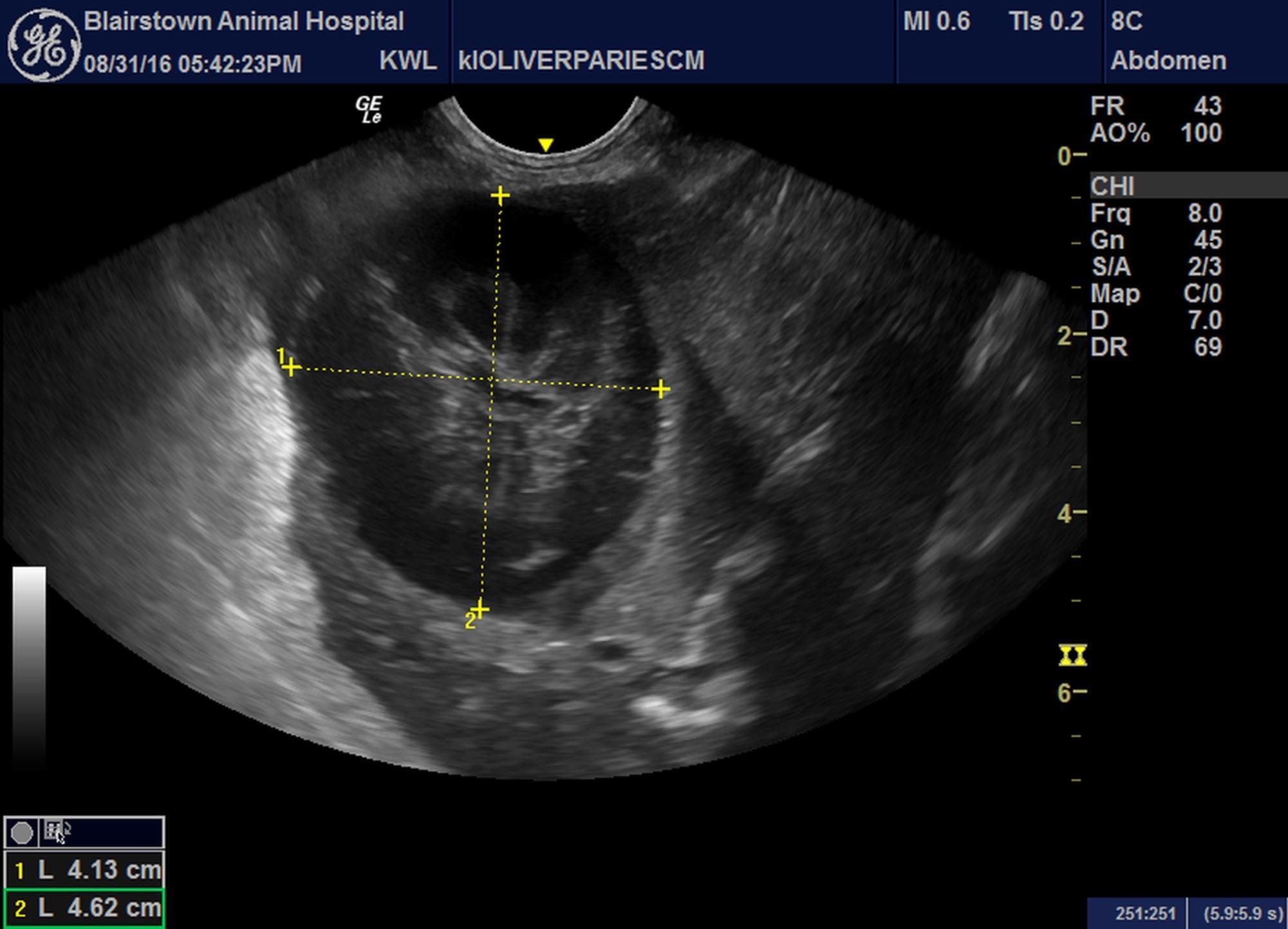
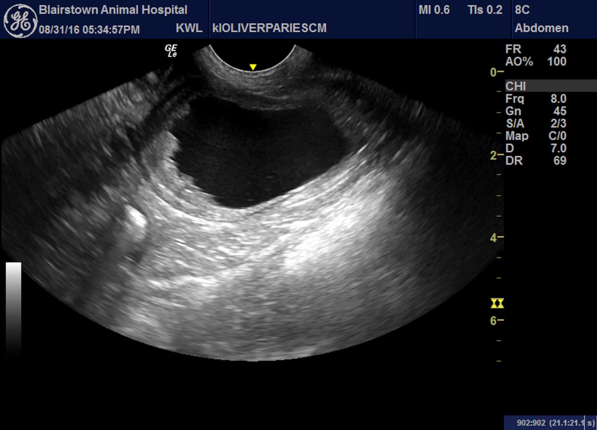
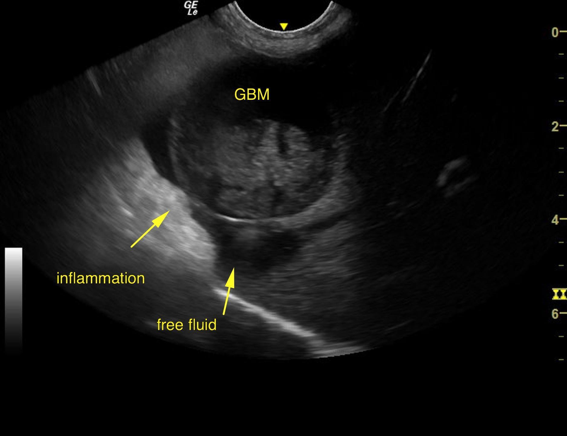
The liver images from right and left intercostal as well as subcostal views revealed subjectively normal liversize, contour, and structure. Parenchymal echogenicity was naturally coarse and hypoechoic to the spleen. Vascular and biliary tracts were of normal volume and no evidence of congestion was noted. The gallbladder presented striating bile and mucocele formation with pericapsular inflammatory pattern. There were minor areas of edema.
The adrenal glands appeared slightly prominent, mildly heterogenic and slightly nodular. No evidence of capsular expansion or invasion into the phrenic veins were noted. No overt suspicion of neoplasia was noted. This is considered likely an age related change or hyperplasia associated with stress or adrenal endocrinopathy with the minimal potential of emerging neoplastic event. The left adrenal gland measured 2.95 x 1.0 cm. The right adrenal gland measured 3.26 x 1.0 cm.
The urinary bladder presented apical polypoid changes that appear to be resectable with resection of the cranial 1/3 of the bladder. This is most consistent with chronic cystitis with potential underlying transitional cell carcinoma. Cytospin of the urine is recommended to assess for pathological cells.