A 1-year-old male neutered Ragdoll cat was with history of chronic eye problems presented for decreased appetite and loose stools for 3 days. Physical exam found patient pyrexic at 104.4 degrees fahrenheit. Suspected enlarged lymph nodes were found on abdominal palpation. Subcutaneous fluids were administered, and patient was discharged with appetite stimulants. Blood chemistry and CBC were within normal limits. FELV/FIV and FIP tests were all negative. Toxoplasma results were normal.
A 1-year-old male neutered Ragdoll cat was with history of chronic eye problems presented for decreased appetite and loose stools for 3 days. Physical exam found patient pyrexic at 104.4 degrees fahrenheit. Suspected enlarged lymph nodes were found on abdominal palpation. Subcutaneous fluids were administered, and patient was discharged with appetite stimulants. Blood chemistry and CBC were within normal limits. FELV/FIV and FIP tests were all negative. Toxoplasma results were normal.
Case Study
FIP in a 1 year old MN Ragdoll cat
Sonographic Differential Diagnosis
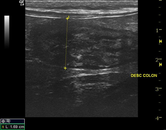
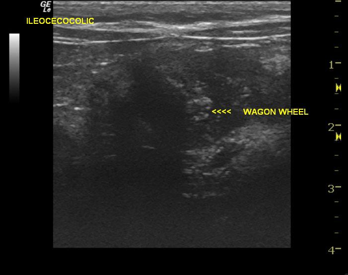
The distal intestinal/colonic mass is suggestive of granulomatous disease (given the age) such as FIP. There is less likely potential for complicated inflammatory disease (resistant bacteria), round cell neoplasia, carcinoma.
Image Interpretation
A mixed hypoechoic intestinal mass is visible with areas of detail loss. Significant mural thickening is noted. The lesions in these images appear isolated and potentially resectable. Neoplastic criteria are met, but complicated inflammatory mural disease may also present in this manner.
DX
Outcome
Patient was sent to referral facility for further evaluation of diarrhea and decreased appetite. On admission, patient was more pyrexic at 104.7, with pink, tacky mucous membranes, and had a gassy abdomen. Patient was placed on intravenous fluid therapy pending an abdominal ultrasound with possible exploratory surgery to follow. Patient underwent exploratory surgery with resection, anastomosis, and biopsies. Biopsy of ileocecal mass revealed severe, chronic-active, ulcerative, pyogranulomatous, transmural enterocecolitis. Results from mesenteric lymph node biopsy, which contained a portion of pancreas attached to it found, necrotizing pyogranulomatous lymphadenitis of both samples; most consistent with FIP. A week post-op, patient was doing poorly, was still pyrexic at 104.2, and experiencing weight loss, however, incision was healing well. Patient was discharged with a course of antibiotics, only to return several days later to be euthanized due to poor quality of life.
Comments
No video is available on this patient.
Clinical Differential Diagnosis
Gastroenteritis (viral, parasitic, protozoal, bacterial), GI foreign body, FIP (despite negative titer), juvenile neoplasia (intestinal lymphoma)
Sampling
Full-thickness surgical biopsy of the ileocecal mass revealed severe, chronic-active, ulcerative, pyogranulomatous, transmural enterocecolitis. Results from mesenteric lymph node biopsy, which contained a portion of pancreas attached to it, found necrotizing pyogranulomatous lymphadenitis of both samples. This is most consistent with FIP.
Patient Information
Clinical Signs
- Anorexia
- Diarrhea
Exam Finding
- Enlarged Lymph Nodes
- Fever
Images
Clinical Signs
- Anorexia
- Diarrhea