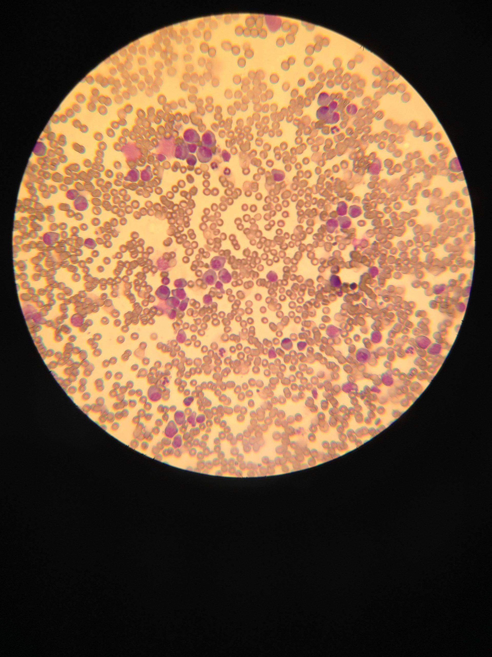
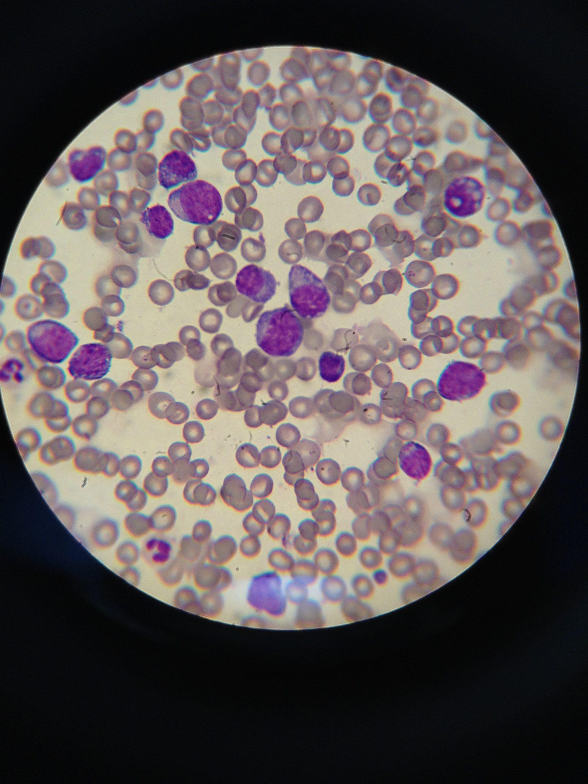
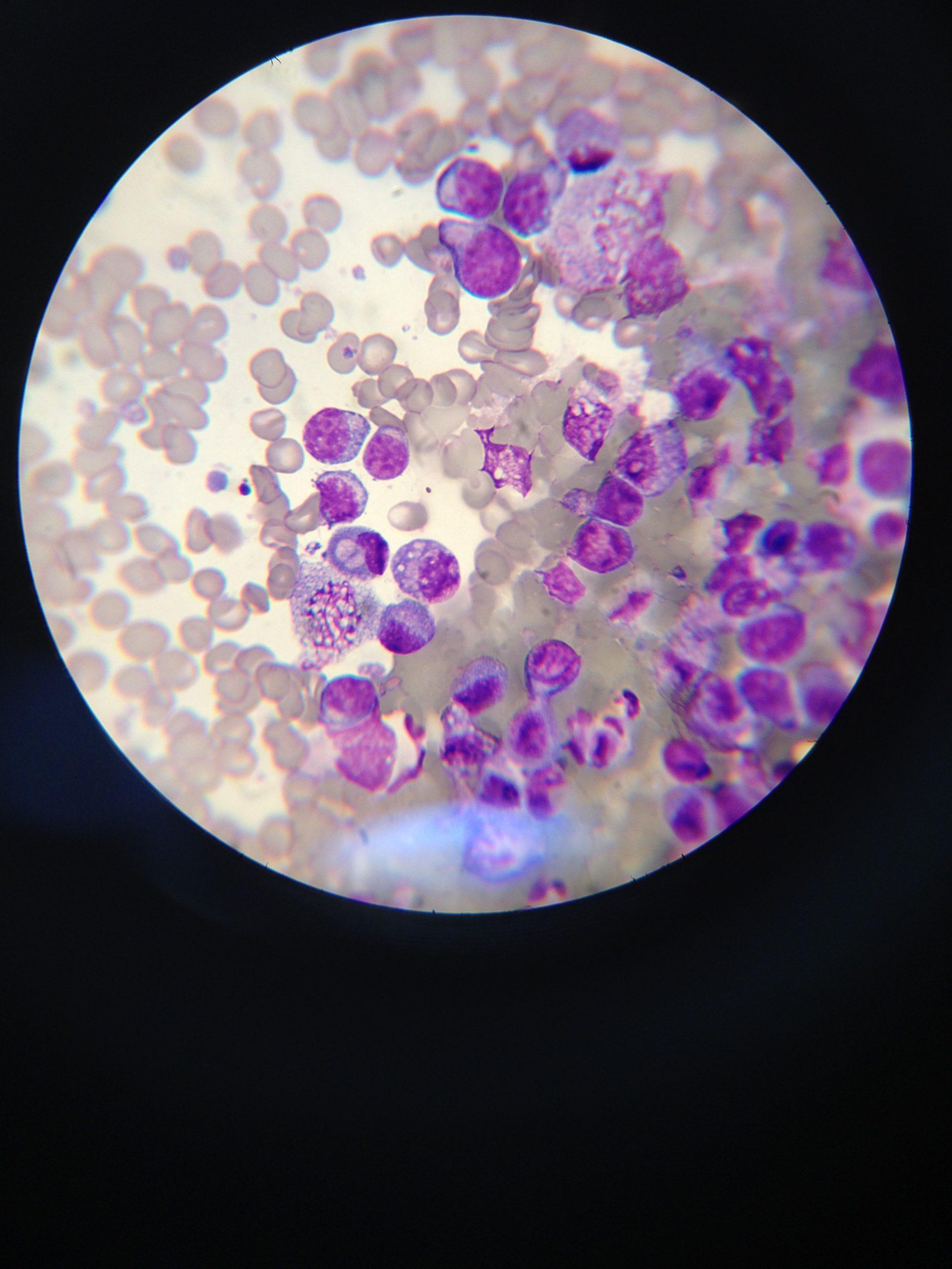
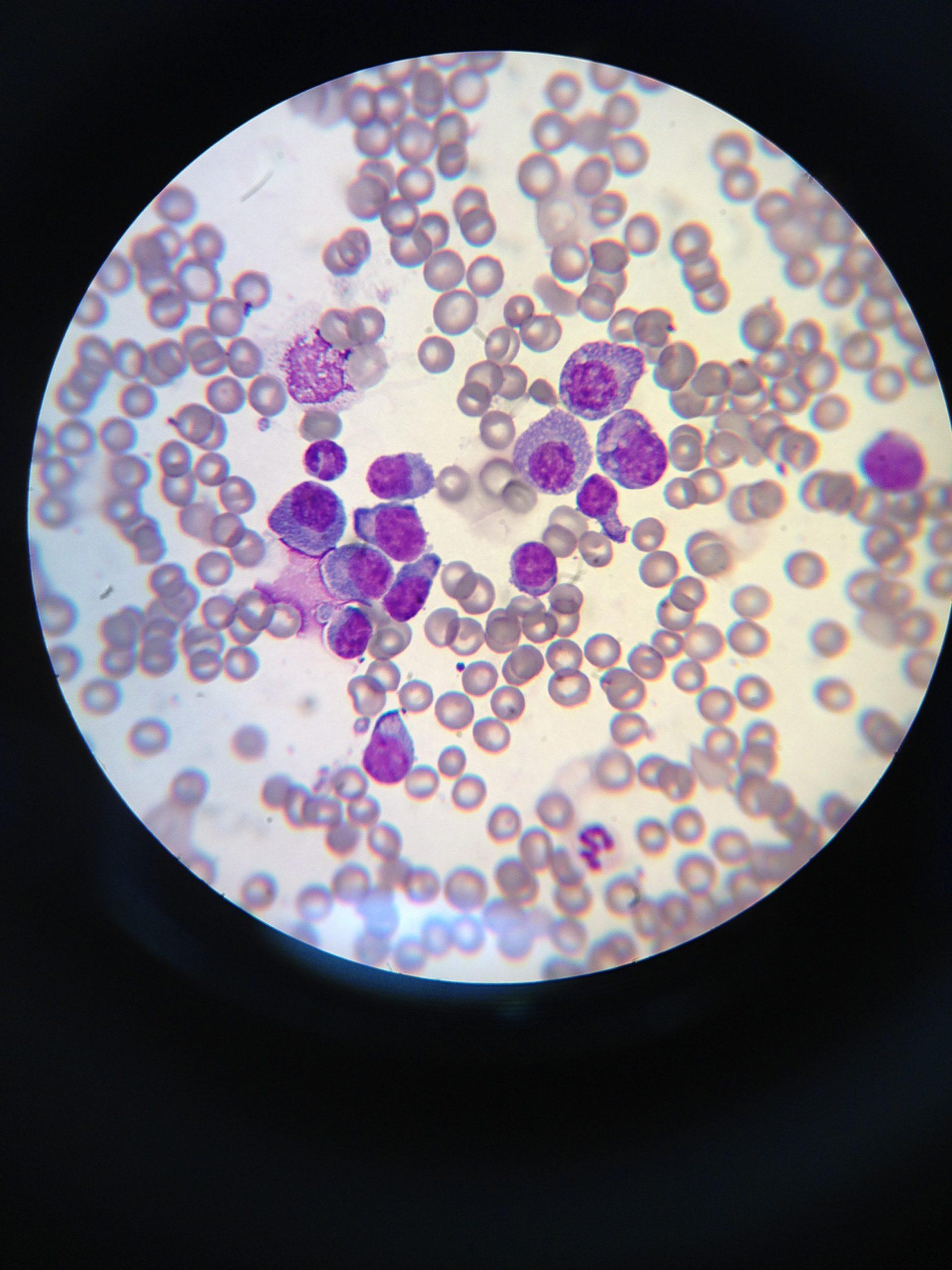
This 11 year old FS Jack Russell Terrier dog has a history of diarrhea of 2 months duration. Painful in caudal abdomen, appetite decreased the past week. On metronidazole and prednisone with no improvement in diarrhea. Ultrasound showed enlarged hypoechoic liver, enlarged hypoechoic mesenteric lymph nodes with reactive fat, trace of abdominal effusion.
CBC: neutrophilia
Chem: BUN, TP, globuolin, chol all low