CT- Tracheal collapse with upper respiratory tract inflammation, rhinitis, tonsillitis, laryngitis in a 14 year old MN West Highland Terrier dog

Case Study

CT- Tracheal collapse with upper respiratory tract inflammation, rhinitis, tonsillitis, laryngitis in a 14 year old MN West Highland Terrier dog

This 14 year old MN West Highland Terrier dog presented with a 2 month history of stertor. 

Physical Exam: audible whistling sound from nose when sniffing

This 14 year old MN West Highland Terrier dog presented with a 2 month history of stertor. 

Physical Exam: audible whistling sound from nose when sniffing

Image Interpretation

CT of the head and thorax, plain and post contrast- There is a mid cervical tracheal collapse. The degree and length of the collapse cannot
be determined since the endotracheal tube and its cuff keeps the tracheal lumen patent.
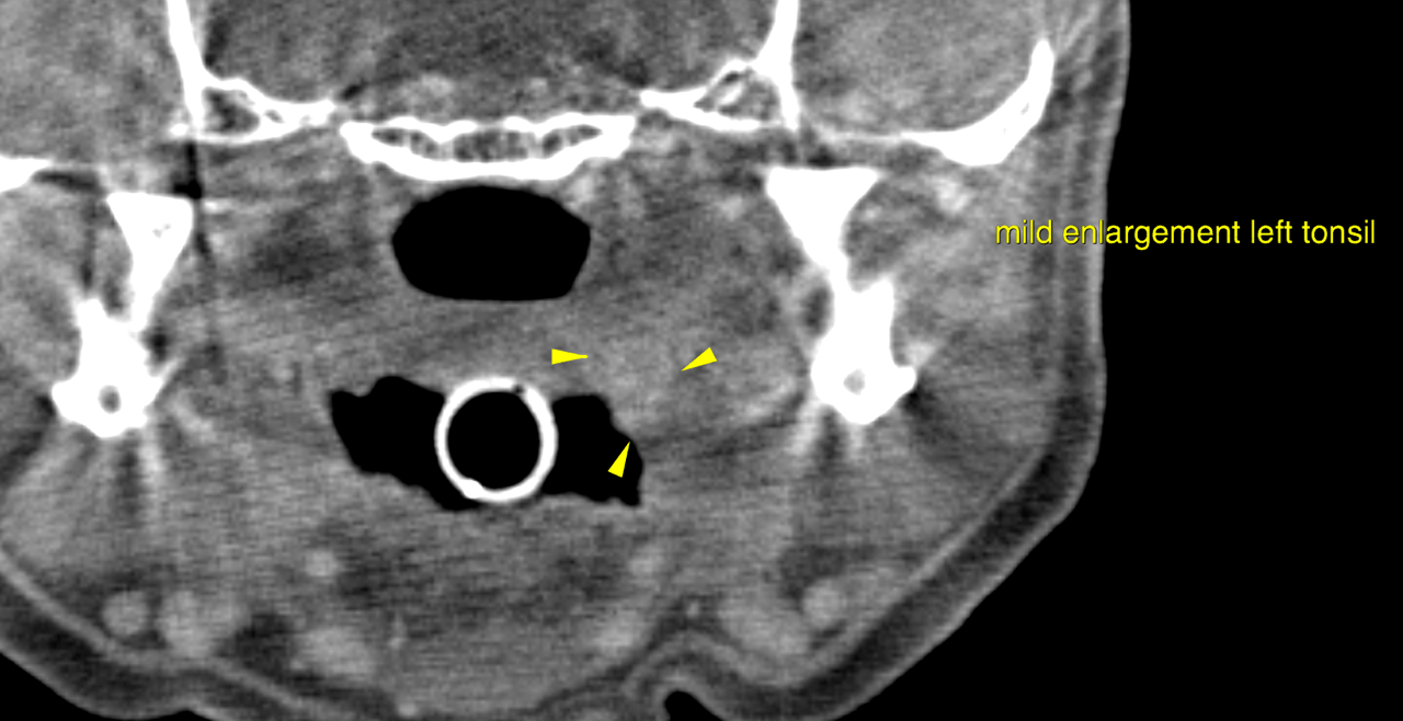
The left tonsil is enlarged and reveals mild protrusion.
The mucosal lining of the laryngeal cartilage reveal mildly irregular thickening.
The soft palate reveals mild symmetrical thickening.
Mild thickening of the nasal cavities’ mucosal linings is noted bilaterally. There is no
mass lesion and no evidence of turbinate destruction. The frontal sinuses are aerated,
there is no abnormal content noted.
Incomplete dentition with loss of multiple teeth and moderate generalized atrophy of
all jaw quadrants is noted.
The pulmonary parenchyma reveals expected geriatric changes. There is no evidence
of parenchymal lung disease or a mass lesion associated with the lung or mediastinal
lymph nodes.
Multifocal mild to moderate degenerative changes are noted along the axial skeleton.
Multifocal osteopenic areas are seen along the cortex of the left humerus, which is at
the very edge of the scan field.

DX

Mid cervical tracheal collapse - Unspecific upper respiratory tract inflammation/infection with mild nondestructive bilateral rhinitis, laryngitis/tracheitis, tonsillitis - Incomplete dentition and age related jaw bone atrophy - Possible multifocal osteopenia left humerus versus artifact

Outcome

Consider rhinoscopy and laryngotracheobronchoscopy with possible sampling for
further structural and functional evaluation of the respiratory tract.
Radiographic evaluation of the left humerus is advised in order to rule out possible
relevant changes here.

Patient Information

Patient Name : Cody Ruda/ALoha
Gender : Male, Neutered
Species : Canine
Type of Imaging : Ultrasound
Status : Complete
Liz Wuz Here : Yes

Clinical Signs

  • Nasal congestion

Images

bildschirmfoto_2016-01-28_um_11

Clinical Signs

  • Nasal congestion
Skip to content