CT- Subacute lung lobe torsion with secondary pleural effusion in a 11 year old MN Chihuahua dog

Case Study

CT- Subacute lung lobe torsion with secondary pleural effusion in a 11 year old MN Chihuahua dog

This 11 year old MN Chihuahua dog presented with pleural effusion

This 11 year old MN Chihuahua dog presented with pleural effusion

Image Interpretation

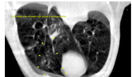
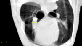
CT of the thorax, plain and post contrast- The main bronchus of the right middle lobe emerges in a ventral direction from the
bifurcation. The bronchus is dilated with an abrupt ending and collapsed lumen 5 mm
after its origin. There is regional lobar opacification and volume loss at this level. Air
filled bronchi are recognized few centimeters distal to the abrupt collapse again. The
distal portion of the right middle lobe is aerated but reveals low volume and is
displaced caudally between the right caudal and accessory lung lobes.
There is moderate bilaterally symmetric pleural effusion.
The remainder of the lung lobes do not reveal abnormalities except for positional
atelectasis.
The mediastinal structures are within normal limits.

DX

subacute lung lobe torsion of the right middle lobe and secondary pleural effusion.

Outcome

Surgical lung lobe resection is recommended.

Patient Information

Patient Name : Bruno Tonn/ Aloha
Gender : Male, Neutered
Species : Canine
Type of Imaging : Ultrasound
Status : Complete
Liz Wuz Here : Yes

Exam Finding

  • Respiratory Distress

Images

screen_shot_2016-03-09_at_12screen_shot_2016-03-09_at_12
Skip to content