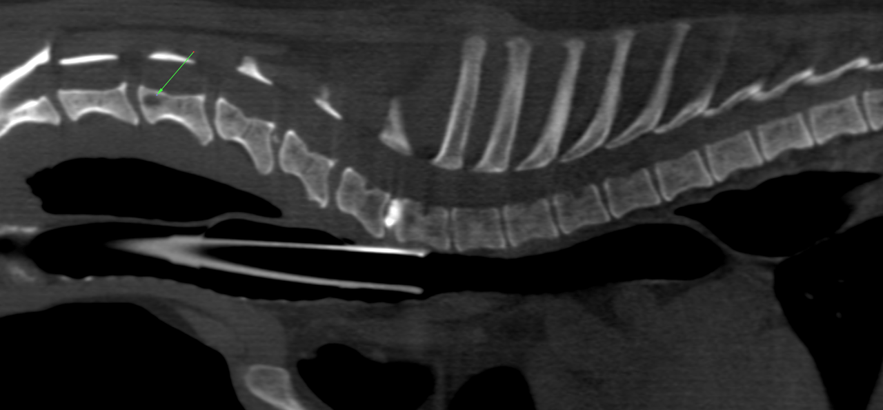
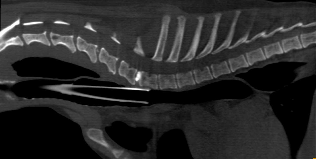
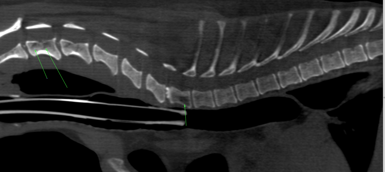
History of sudden onset lameness approximately 7/23/15 after falling off steps. She stopped using the right front on 7/25/15 altogether.
History of sudden onset lameness approximately 7/23/15 after falling off steps. She stopped using the right front on 7/25/15 altogether.
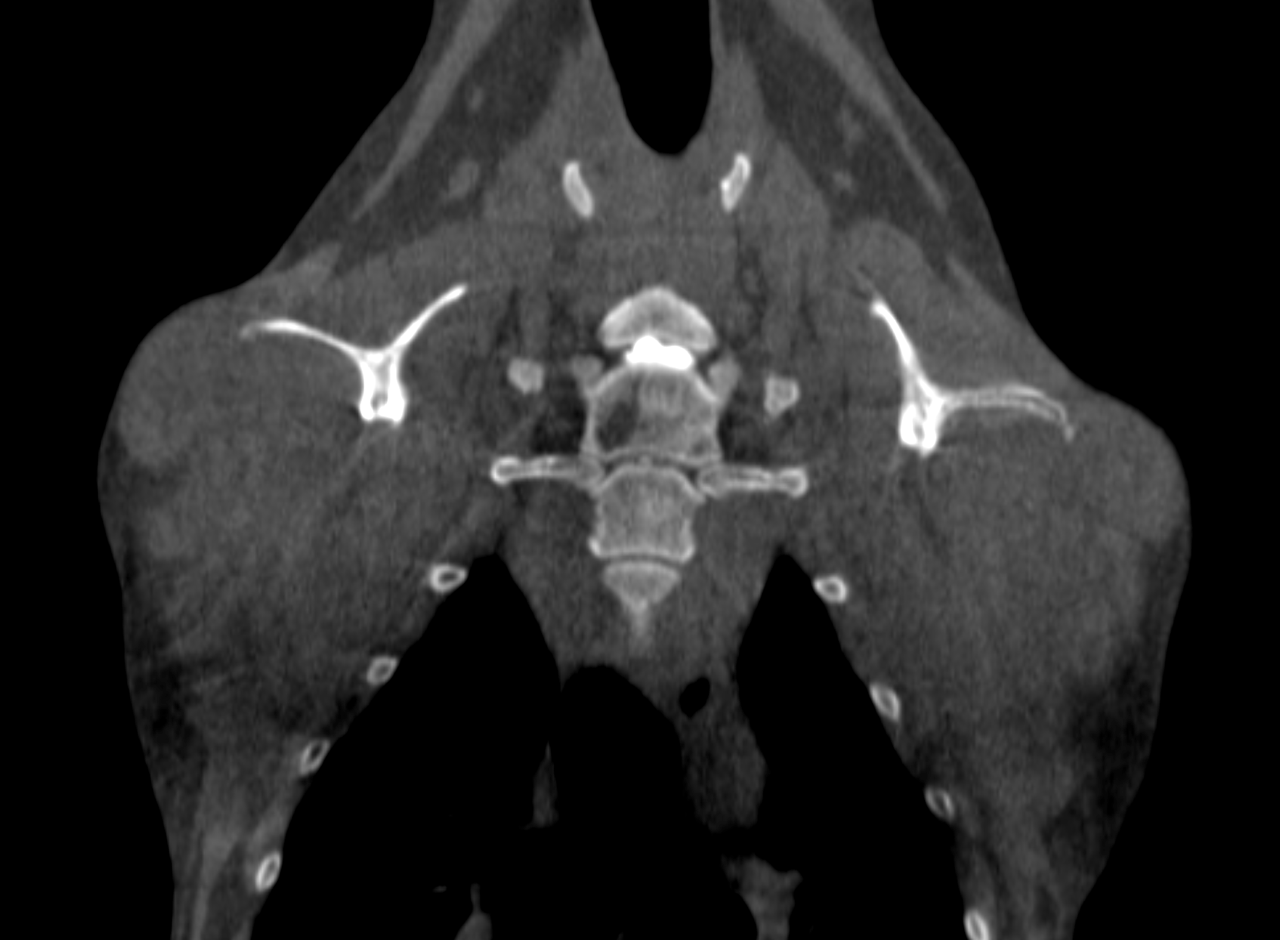
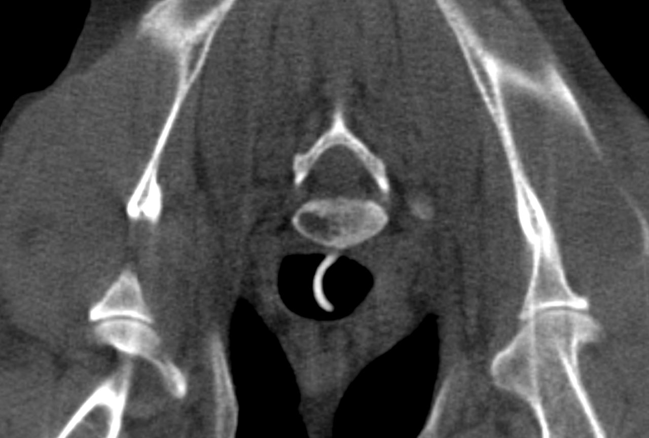
Physical Exam: Alert and responsive. Normal heart and lung auscultation, no pain noted on abdominal palpation and normal lymph nodes. There is normal placing but the gait is showing no shoulder movement and circumduction on gait. Both shoulders were subluxated and replaced in the exam room; this gave dog much more mobility. The left thoracic limb was looser after replacement and withdrawal was present. Some pain at the right bicipital tendon insertion which disappeared with massage and range of motion. Withdrawal was noted after a few tries. No neck pain.