Skip to content

CT – Right auricular mass with scant pericardial effusion in a 12 year old FS dog

Case Study

CT – Right auricular mass with scant pericardial effusion in a 12 year old FS dog

This 12 year old FS dog has a right atrial mass noted on ultrasound; previous pericardial effusion

This 12 year old FS dog has a right atrial mass noted on ultrasound; previous pericardial effusion

Image Interpretation

CT of the thorax – 

A non-uniformly enhancing multinodular mass of 2.8 cm diameter is seen at the right auricular appendage causing a mass effect onto the right atrium. Stranding of the pericardial fat is noted.  The caudal aspect of the right cranial lung lobe presents a homogeneous soft tissue attenuating area and reduced volume. The margins of the liver are rounded and the liver is prominent. The gastric axis is rotated caudally.

DX

Cardiac mass at the right auricular appendage with scant pericardial effusion and mass effect onto the right atrium

Outcome

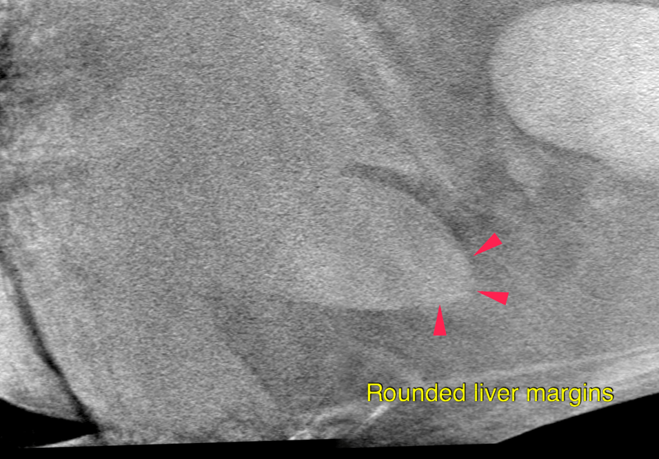
Also moderate generalized hepatomegaly; No evidence of metastatic spread to the lung parenchyma, mediastinal lymph nodes and included parts of the spleen (only small part included).
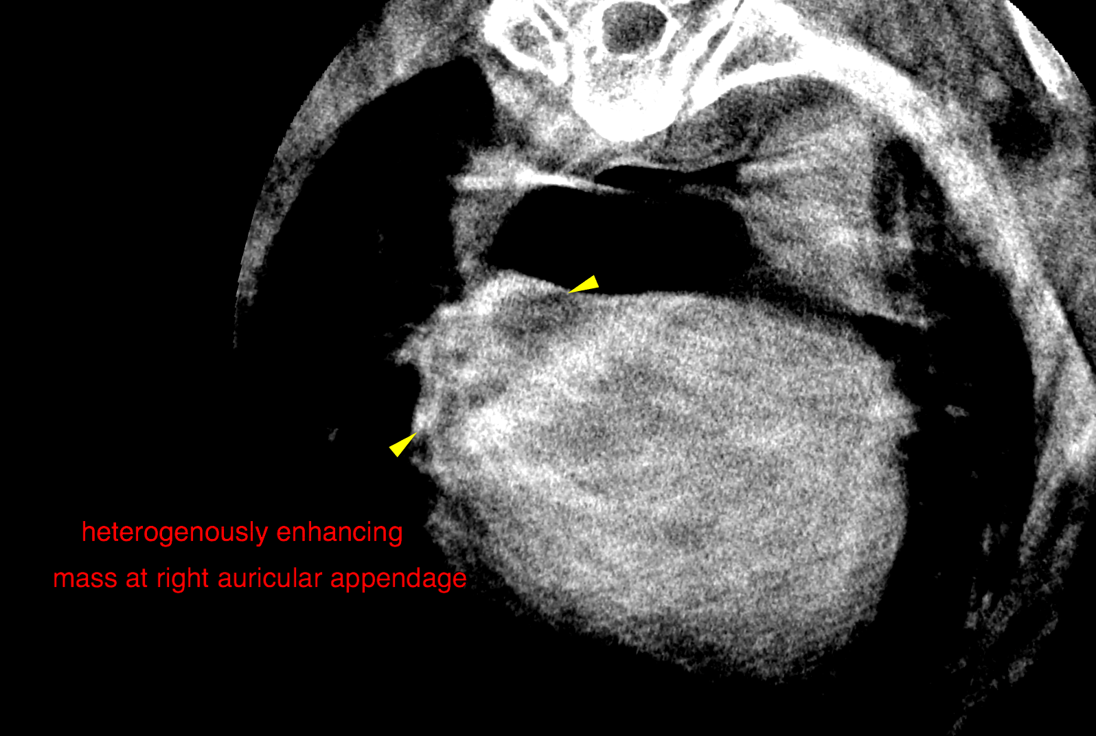
Right atrial hemangiosarcoma is the main differential diagnosis here. Other heart base
masses, such as apudoma (formerly known as chemodectoma), ectopic
thyroid/parathyroid carcinoma or lymphosarcoma/histiocytic sarcomas are less likely.
The hepatomegaly can be caused either by congestion due to the possible mass lesion
within the right atrium or is due to a diffuse infiltrative hepatopathy. Possible
differential diagnosis for hepatopathy are fatty infiltration, steroid induced
hepatopathy, vacuolar hepatopathy, hepatitis, diffuse neoplastic infiltrate.

Patient Information

Patient Name : Lucy Schifano
Gender : Female, Spayed
Species : Canine
Type of Imaging : Ultrasound
Liz Wuz Here : Yes
Code : 16_00111

Exam Finding

  • Masses

Images

bildschirmfoto_2016-08-23_um_13bildschirmfoto_2016-08-23_um_11bildschirmfoto_2016-08-23_um_11