History of difficulty walking for the past several months. Physical exam showed grade 4/6 cardiac murmur.
History of difficulty walking for the past several months. Physical exam showed grade 4/6 cardiac murmur.
History of difficulty walking for the past several months. Physical exam showed grade 4/6 cardiac murmur.
History of difficulty walking for the past several months. Physical exam showed grade 4/6 cardiac murmur.
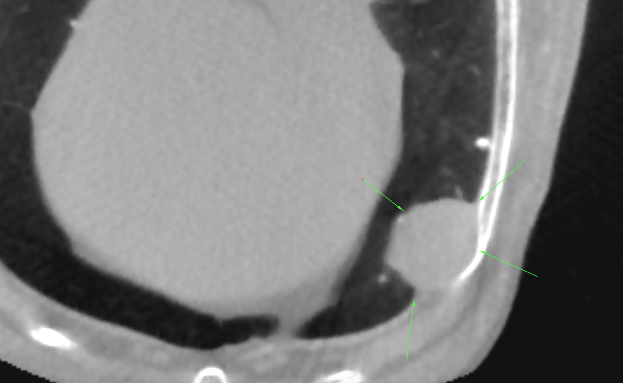
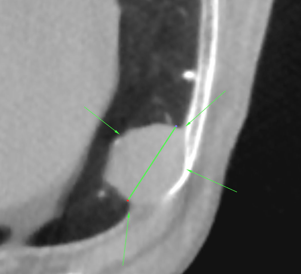
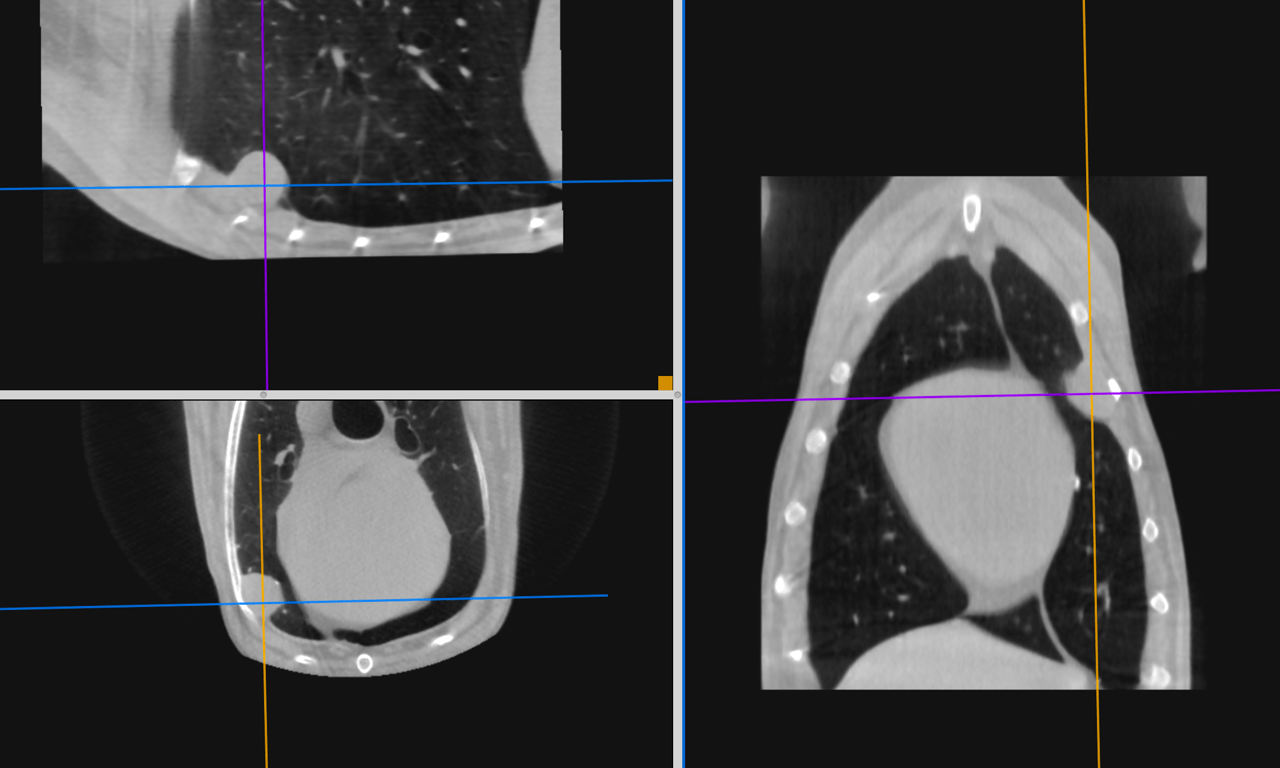
CT of the thorax, plain and post contrast series – There was a soft tissue attenuating, low enhancing ovoid mass lesion measuring approximately 1.5 cm in diameter within the left pleural cavity adjacent to the fourth rib and left cranial lung lobe. The lesion appeared to displace the lung lobe medially but was irregularly contoured to the lung parenchyma. The mass had narrow angles to the chest wall resembling an extrapleural/pleural sign. There was a mass effect causing pressure atrophy of the fourth rib localized to its ventral third. Minor atelectatic and fibrotic changes were seen within the lung parenchyma adjacent to the mass.
There were multifocal small mineralized foci throughout the lungs and pleura consistent with pleural plugs/pulmonary osteomata.
The mediastinal lymph nodes were within normal limits.
Specific malignancy criteria are not evident so principally benign and malignant tumors have to be considered.
Sampling is strongly recommended which should ideally be done under ultrasound guidance within the 3rd or 4th intercostal space using a parasternal approach.
Conditions for ultrasound guided sampling are ideal since the mass is superficial immediately adjacent to the chest wall. Avoid puncture of the adjacent lung. But also note that the risk of causing a clinical iatrogenic pneumothorax in case of puncture of the lung is very low in dogs.