CT – Perianal mass with severe generalized lymphadenomegaly in a 7 year old MN Rottweiller mix

Case Study

CT – Perianal mass with severe generalized lymphadenomegaly in a 7 year old MN Rottweiller mix

This 7 year old MN Rottweiller mix presented with right-sided perianal swelling; cytology  report adenocarcinoma. Extensive mass palpated; CT to determine extent of mass, lymph node involvement and presence of metastases.

This 7 year old MN Rottweiller mix presented with right-sided perianal swelling; cytology  report adenocarcinoma. Extensive mass palpated; CT to determine extent of mass, lymph node involvement and presence of metastases.

Image Interpretation

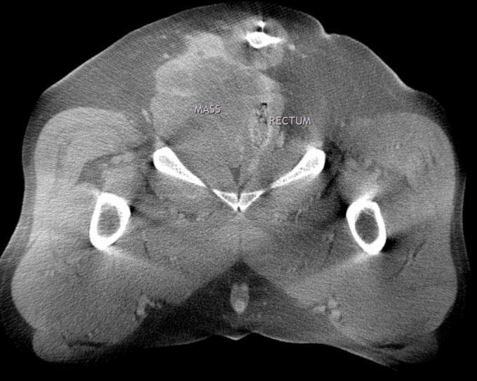
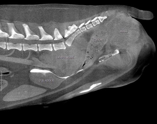
An irregular shaped and ill-defined mass is seen in the right perianal region. The mass lesion measures approximately 8,5cm in the longitudinal axis, 7,6cm in the transverse axis and 7,0cm in the lateral axis. The mass is adjacent to the dorsal border of the right ischium and the ischial symphysis, displaces the rectum and pelvic urethra to the left side and partially compromise their lumen. Tail and coccygeal vertebrae are dorsally displaced. The mass presents heterogeneous soft tissue attenuation with marked heterogeneous contrast uptake with multiple amorphous, coalescing, non-enhancing areas within the mass (tumoral necrosis).

Severe adenomegaly of all hypogastric and medial iliac lymph nodes is moted. The lymph nodes measure approximately 9,5cm in the longitudinal axis, 4,9cm in the transverse axis and 5,6cm in the lateral axis. The lymph nodes present a mass effect on the iliopsoas muscle bilaterally, and are adjacent to the ventral aspect of L6, L7 and sacrum. Ventral displacement of the terminal caudal vena cava, iliac veins (partially obliterating their lumen), terminal aorta, external and internal iliac arteries, sacral median artery, caudal aspect of urinary bladder, urethra and prostate is noted. The descending colon is displaced ventrally and to the right.


DX

Right perianal mass meeting neoplastic criteria with severe generalized lymphadenomegaly

Outcome

The CT findings are consistent with the history of a right anal sac carcinoma and compatible with metastatic spread to the hypogastric and medial iliac lymph nodes. Surgical resection of the anal sac mass and lymph nodes may be limited by their size and restricted by the bony anatomy of the pelvis.Peripheral tissue infiltration cannot be ruled out.

Patient Information

Patient Name : Ridley Choate
Gender : Male, Neutered
Species : Canine
Type of Imaging : Ultrasound
Status : Complete
Liz Wuz Here : Yes
Code : 16-00165

Clinical Signs

  • Swelling

Exam Finding

  • Masses

Images

ridley_01ridley_02

Clinical Signs

  • Swelling
Skip to content