CT – Odontogenic or soft tissue neoplasia at area of canine tooth in a 8 year old FS Jack Russell Terrier dog

Case Study

CT – Odontogenic or soft tissue neoplasia at area of canine tooth in a 8 year old FS Jack Russell Terrier dog

This 8 year old FS Jack Russell Terrier dog presented with ameloblastoma at the right lower canine. 

This 8 year old FS Jack Russell Terrier dog presented with ameloblastoma at the right lower canine. 

Image Interpretation

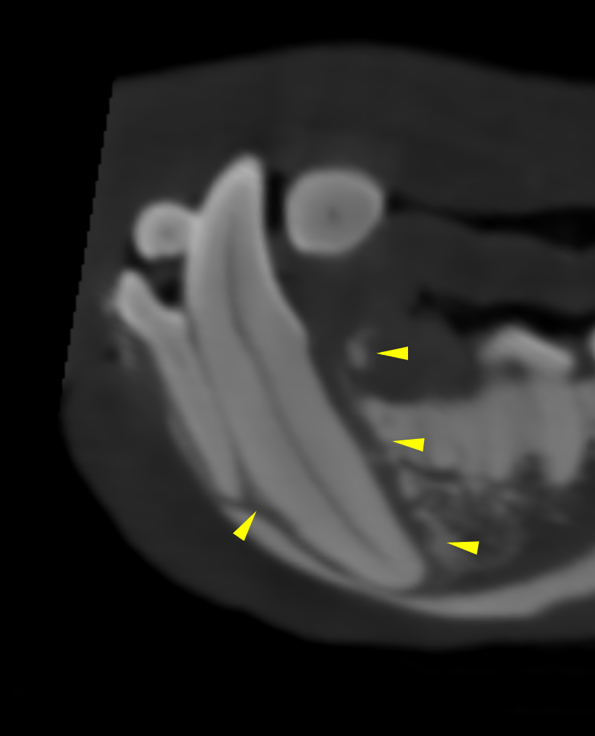
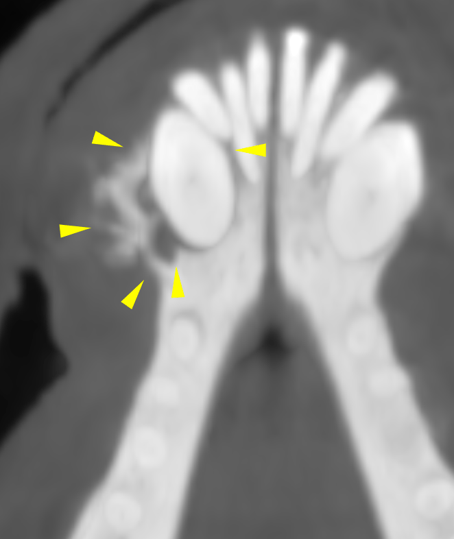
CT of the head, plain and post contrast – The computed tomography reveals a soft tissue mass with heterogenous contrast
uptake and punched out osteolytic lesions centered around the right lower canine tooth
(Triadan 404). The lesion expands the periodontal ligament, lamina dura and overlying
cortex emphasizing the lateral and caudal aspect of the alveole. A moderate amount of
periosteal new bone is arranged in a sunburst pattern lateral to the alveolar bone. The
osteolysis does not extended beyond the mandibulary symphysis. The neighbouring
teeth and alveolar bone are within normal limits.
The lymph nodes of the head are within normal limits.

DX

The computed tomographic findings match the history of a malignant odontogenic or soft tissue neoplasia such as ameloblastoma (or acanthomatous epuli, squamous cell carcinoma) at Triadan 404.

Outcome

Partial hemimandibulectomy should be considered. With the minimally required safety
margins of 1 cm the mandibular ramus should be removed covering for all right lower
incisors and premolar 1 & 2 (Triadan 405 & 406) at least.

Patient Information

Patient Name : Abbie Young/Aloha
Gender : Female, Spayed
Species : Canine
Type of Imaging : Ultrasound
Status : Complete
Liz Wuz Here : Yes

Exam Finding

  • Doughy Abdomen

Images

bildschirmfoto_2016-02-03_um_18bildschirmfoto_2016-02-03_um_18bildschirmfoto_2016-02-03_um_19
Skip to content