CT – Non-specific recurrent pleural effusion in a 9 year old MN Rat Terrier

Case Study

CT – Non-specific recurrent pleural effusion in a 9 year old MN Rat Terrier

This 9 year old MN Rat Terrier dog has a history of recurrent pleural effusion consisting of modified transudate with lymphocytes

This 9 year old MN Rat Terrier dog has a history of recurrent pleural effusion consisting of modified transudate with lymphocytes

Image Interpretation

CT of the thorax, plain and post contrast – 

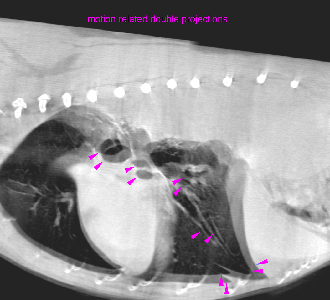
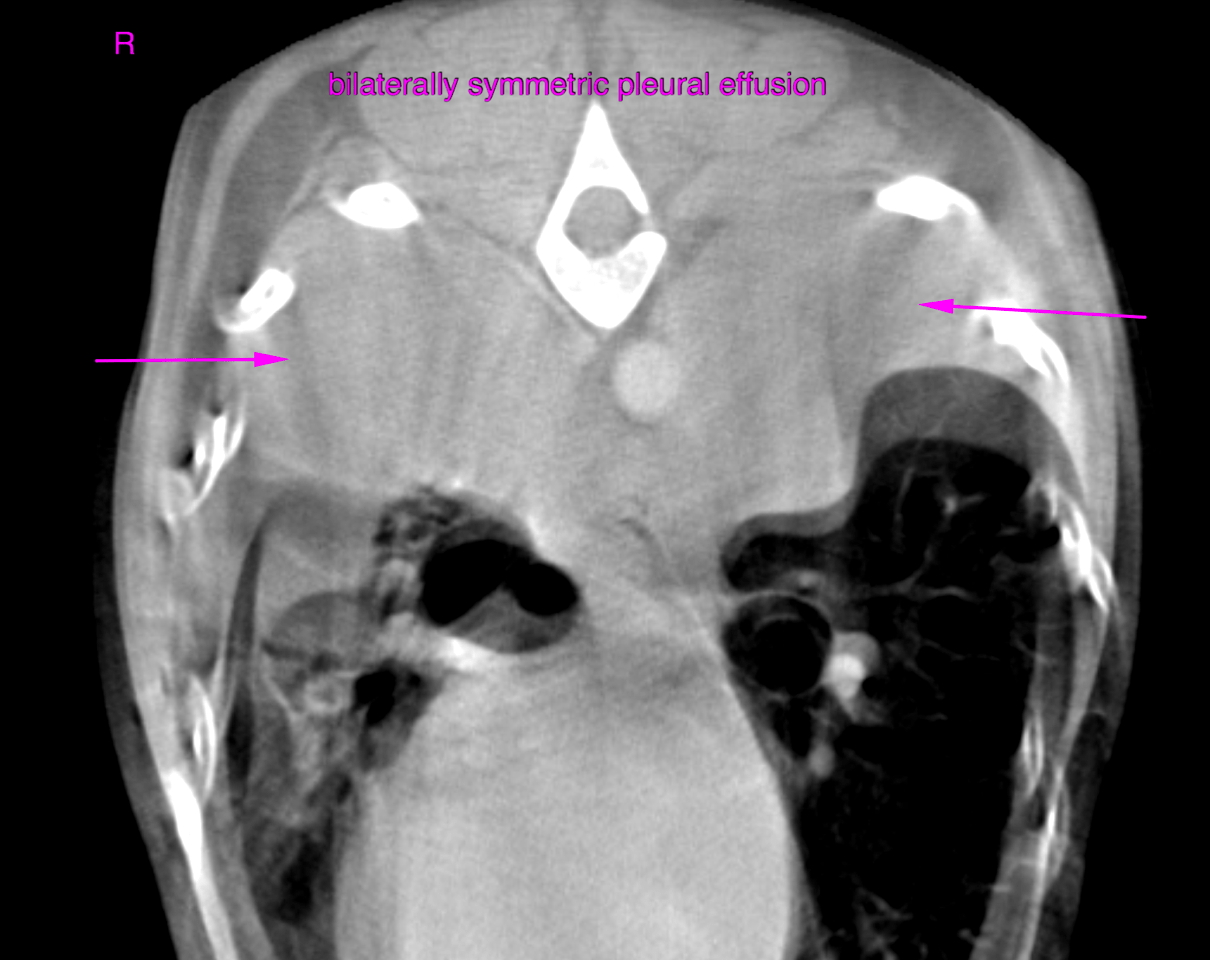
Moderate bilateral, non-compartimentalized pleural effusion is noted in the gravity dependent portions of the pleural cavities. The lung parenchyma and the bronchial tree present no abnormalities except for peripheral atelectasis.  The pulmonary vasculature is within normal limits. There is no evidence of lung lobe torsion. The mediastinum and mediastinal lymph nodes are within normal limits. The heart is within normal limits, there is no specific cardiac chamber or major vessel enlargement and no evidence of congestive heart failure. There is no pericardial effusion.

DX

Non-specific moderate bilaterally symmetric pleural effusion without evidence of cardiac, pulmonary neopasia, pulmonary parenchymal or airway disease.

Outcome

Consider idiopathic chylous/pseudochylous effusion. Primary pleural neoplasia is a possible differential diagnosis with lower potential.

Patient Information

Patient Name : Copper Stewart/Aloha
Gender : Male, Neutered
Species : Canine
Type of Imaging : Ultrasound
Status : Complete
Liz Wuz Here : Yes
Code : 16_00031

Images

bildschirmfoto_2016-06-07_um_13bildschirmfoto_2016-06-07_um_13