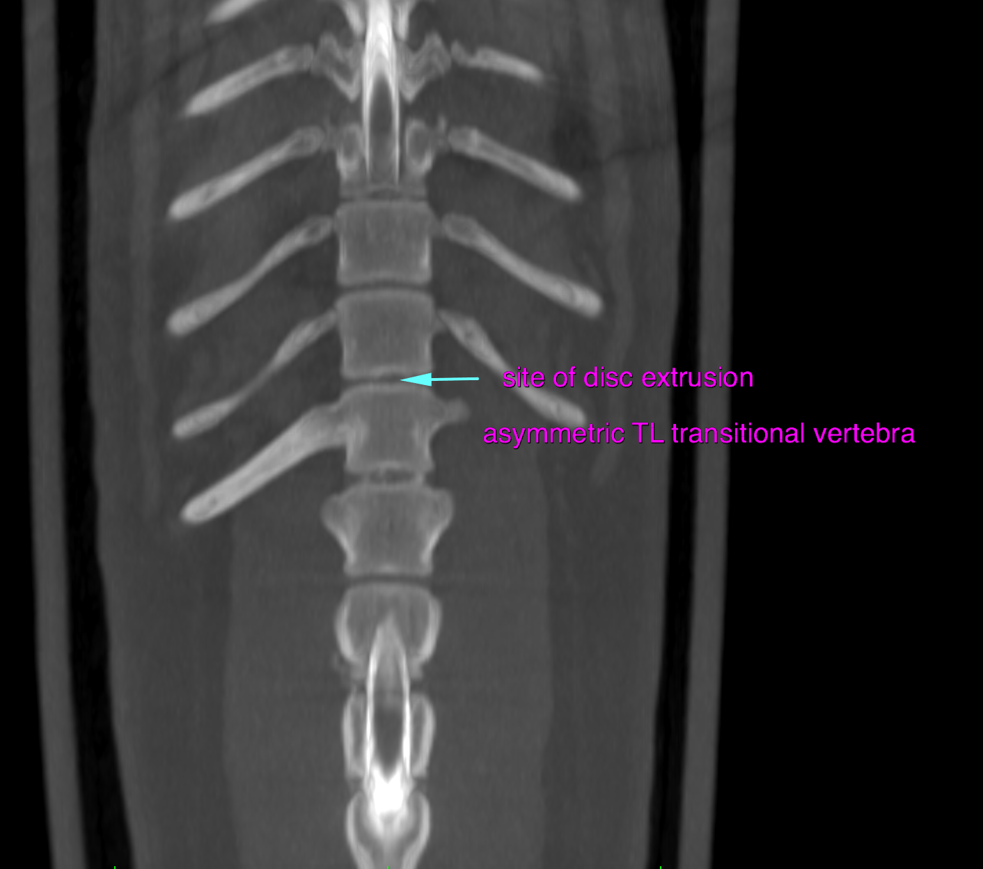
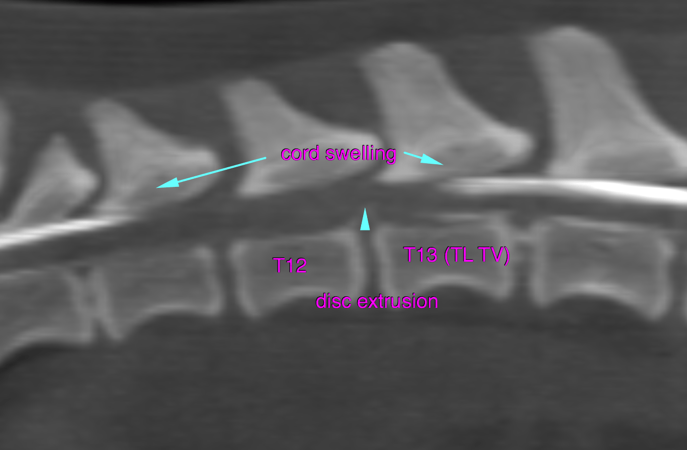
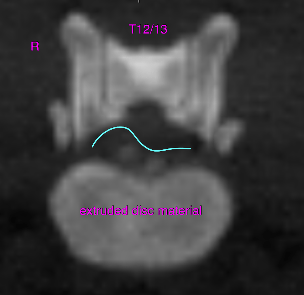
This 4 year old MN Lhasa Apso presented for pelvic limb ataxia progressing to paresis. CP absent, reflexes +3, panniculus present until T-L junction. Suspect IVDD
This 4 year old MN Lhasa Apso presented for pelvic limb ataxia progressing to paresis. CP absent, reflexes +3, panniculus present until T-L junction. Suspect IVDD