CT – Congenital transitional lumbosacral vertebra, acquired lumbosacral disc protrusion, and periodontitis in a 13 year old FS Rat Terrier dog.

Case Study

CT – Congenital transitional lumbosacral vertebra, acquired lumbosacral disc protrusion, and periodontitis in a 13 year old FS Rat Terrier dog.

The patient is a 13 year old FS Rat Terrier dog. An abdominal ultrasound was submitted for the patient last week. Pet is painful/sensitive in thoracic spine and reluctant to eat.


The patient is a 13 year old FS Rat Terrier dog. An abdominal ultrasound was submitted for the patient last week. Pet is painful/sensitive in thoracic spine and reluctant to eat.


Image Interpretation

CT of the spine and mouth – There is a congenital asymmetric lumbosacral transitional vertebra, which is likely to
be an incidental finding here.
Developing lumbosacral, L4/5 and L5/6 spondylosis is noted.
There is a moderate protrusion of the lumbosacral disc.
Moderate positional atelectasis is noted within the dependent portions of the lung.
Dentition:
The dentition is complete.
Moderate bone atrophy is developed in the incisor part of the maxilla and mandible
emphasizing the left upper quadrant.
Mild generalized horizontal alveolar bone atrophy is noted for all quadrants.
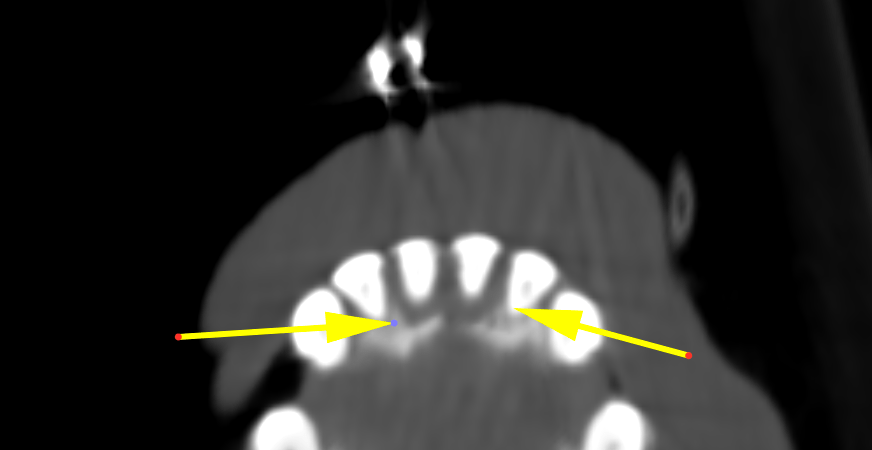
The upper canine teeth reveal moderate resorptive changes and developing age related
ankylosis.
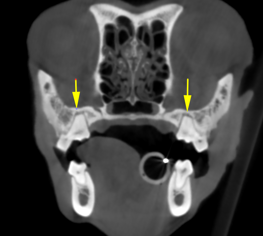
Mild widening of the periodontal ligament with reduced delineation of the lamina dura is noted in the alveoles of the upper P4, M1 and M2 bilaterally. Similar changes are
noted surrounding the left lower second incisor.
Plaque like coats are noted at the neck of the upper canine teeth and M1 bilaterally
(buccal surface).
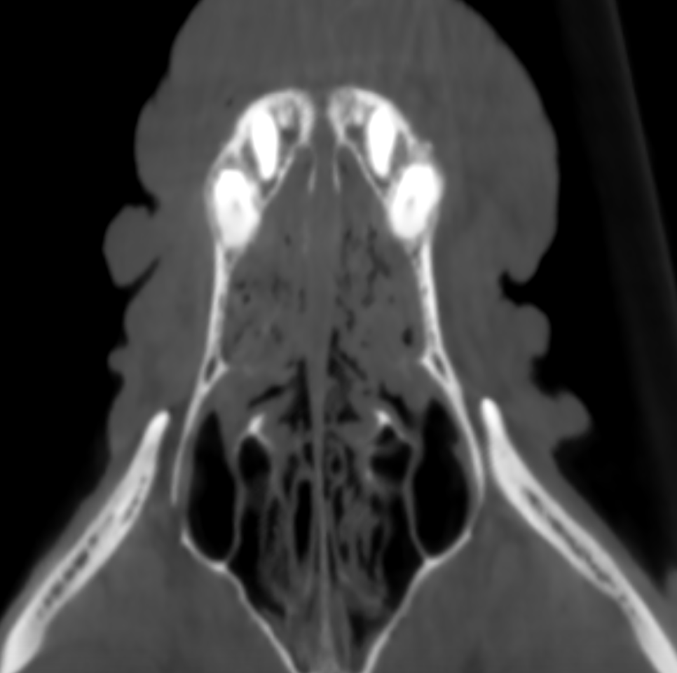
Mild swelling of the mucosal lining of the nasal turbinates is noted for the rostral third
of both nasal cavities.

DX

congenital transitional lumbosacral vertebra and acquired moderate lumbosacral disc protrusion. The clinical significance is questionable especially in a small breed dog. The dentition is complete, the occlusion unremarkable. Chronic periodontitis.

Outcome

Overall moderate age related
changes are developed in all quadrants. There are signs of mild chronic periodontal
inflammation at the carnassial and molar teeth in both upper jaws. Furthermore, the
second lower left incisor and the maxillary incisors reveal moderate chronic
periodontitis.
The mild mucosal thickening within the nasal cavities may be a normal finding within
the limits of the nasal cycle or a consequence of mild unspecific non-destructive
rhinitis.
There is no oro-nasal fistula.

Patient Information

Patient Name : Alli Santora
Gender : Female, Spayed
Species : Canine
Type of Imaging : Ultrasound
Status : Complete
Liz Wuz Here : Yes

Clinical Signs

  • Anorexia
  • Pain

Images

bildschirmfoto_2015-12-08_um_12bildschirmfoto_2015-12-08_um_11bildschirmfoto_2015-12-08_um_11

Clinical Signs

  • Anorexia
  • Pain
Skip to content