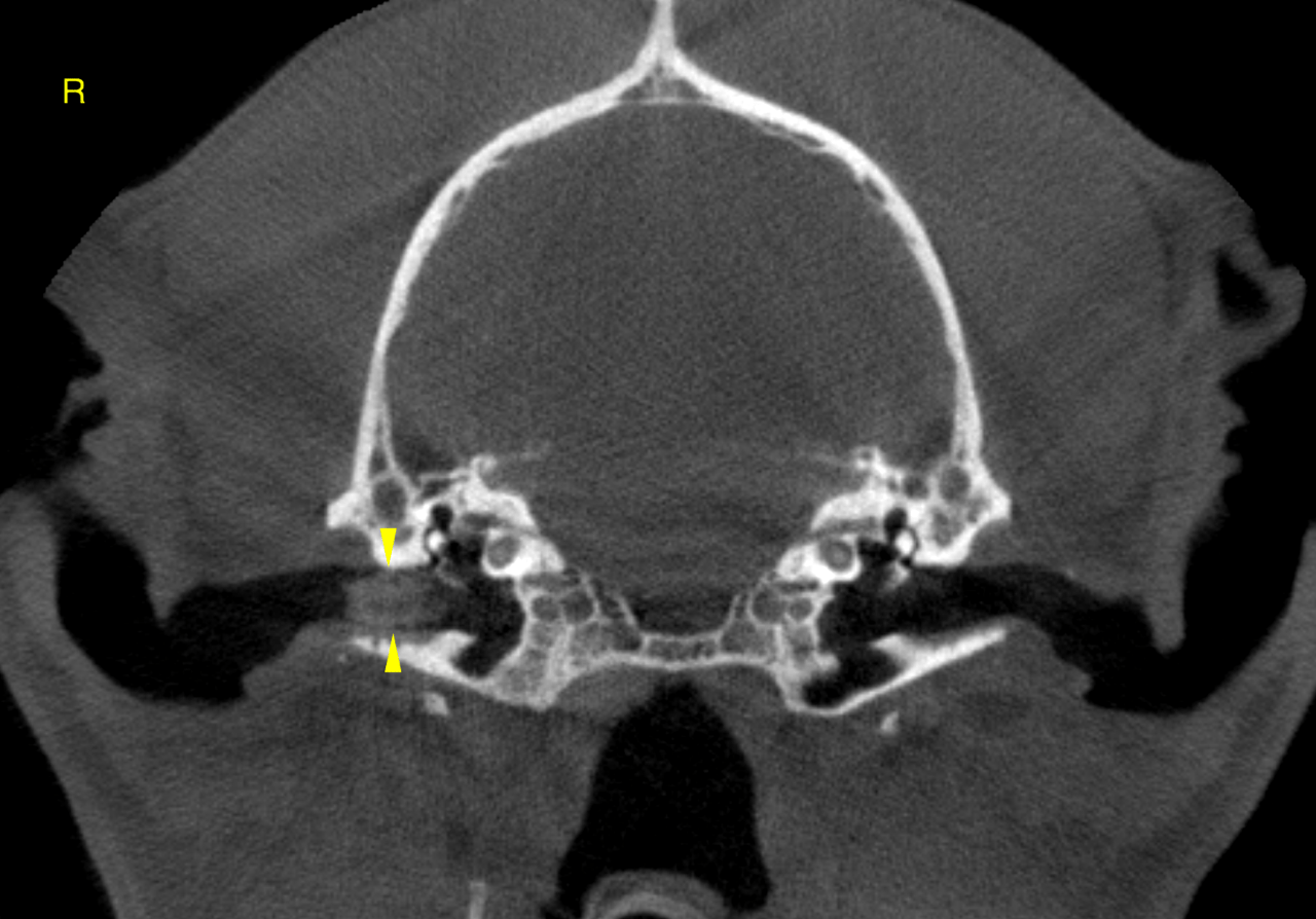
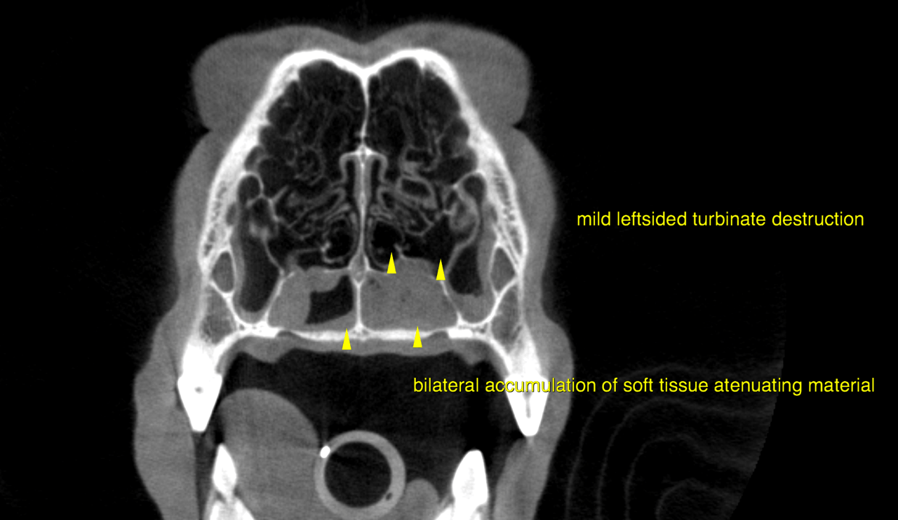
This 4 year old FS Doberman Pinscher dog presented with acute onset nasal discharge. Previous diagnosis of EOS meningitis 2 weeks ag.
Physical exam: purulent blateral nasal discharge
CBC/Chem: wnl
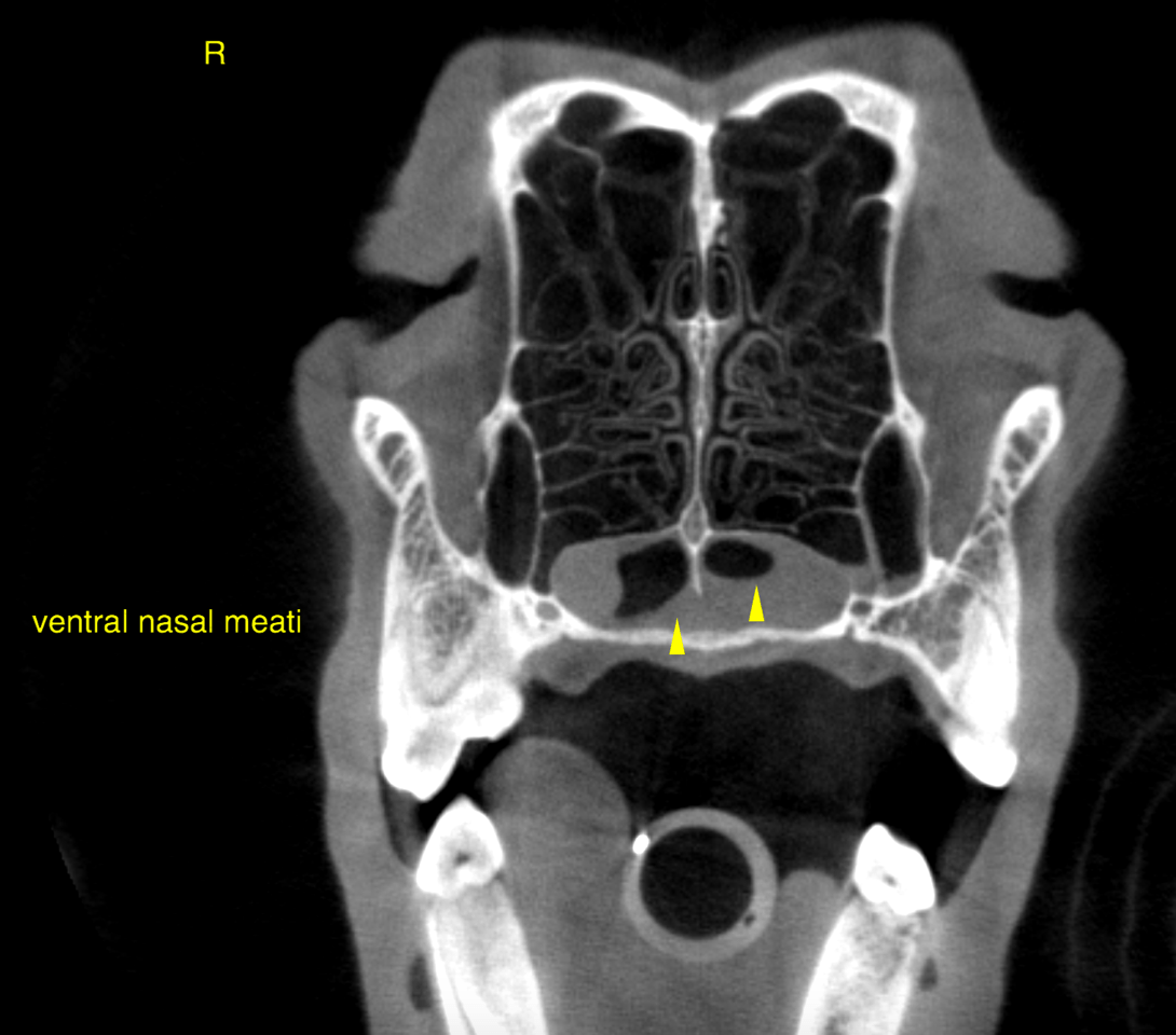
This 4 year old FS Doberman Pinscher dog presented with acute onset nasal discharge. Previous diagnosis of EOS meningitis 2 weeks ag.
Physical exam: purulent blateral nasal discharge
CBC/Chem: wnl