CT of the head and abdomen –
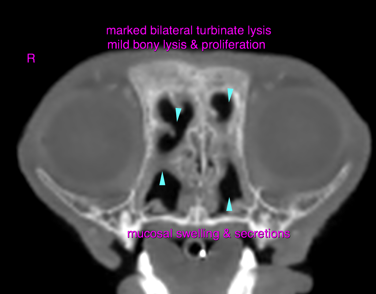
Head: The computed tomography of the head reveals marked bilateral turbinate lysis. The mucosal lining of the remaining turbinates and nasal bones presents moderate swelling. Multifocal small permeative osteolysis and mild osteoproliferations are noted. The cribriform plate is intact. A moderate amount of soft tissue attenuating secretions are seen within both nasal cavities emphasizing the ventral nasal meati as well as the nasal fundus. The sphenoidal sinuses are occupied with soft tissue attenuating material as well. The right frontal sinus is rudimentary which is common in brachycephalic animals and is occupied by soft tissue attenuating material.
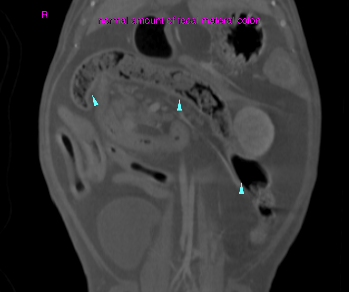
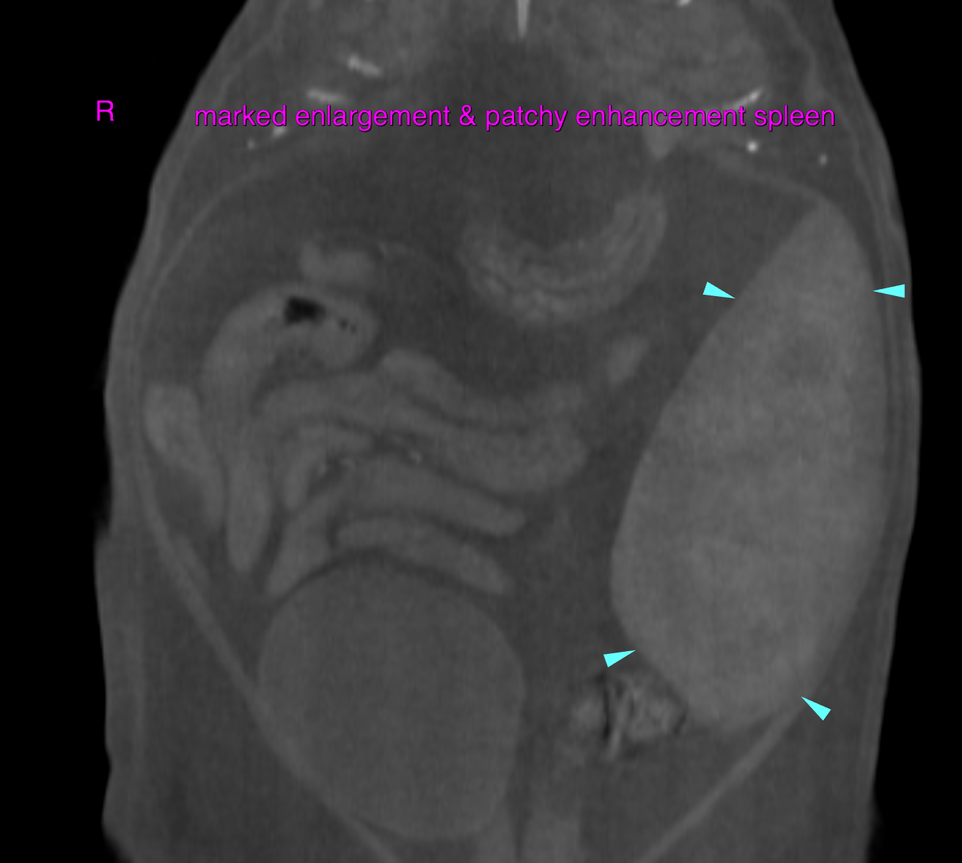
Abdomen:
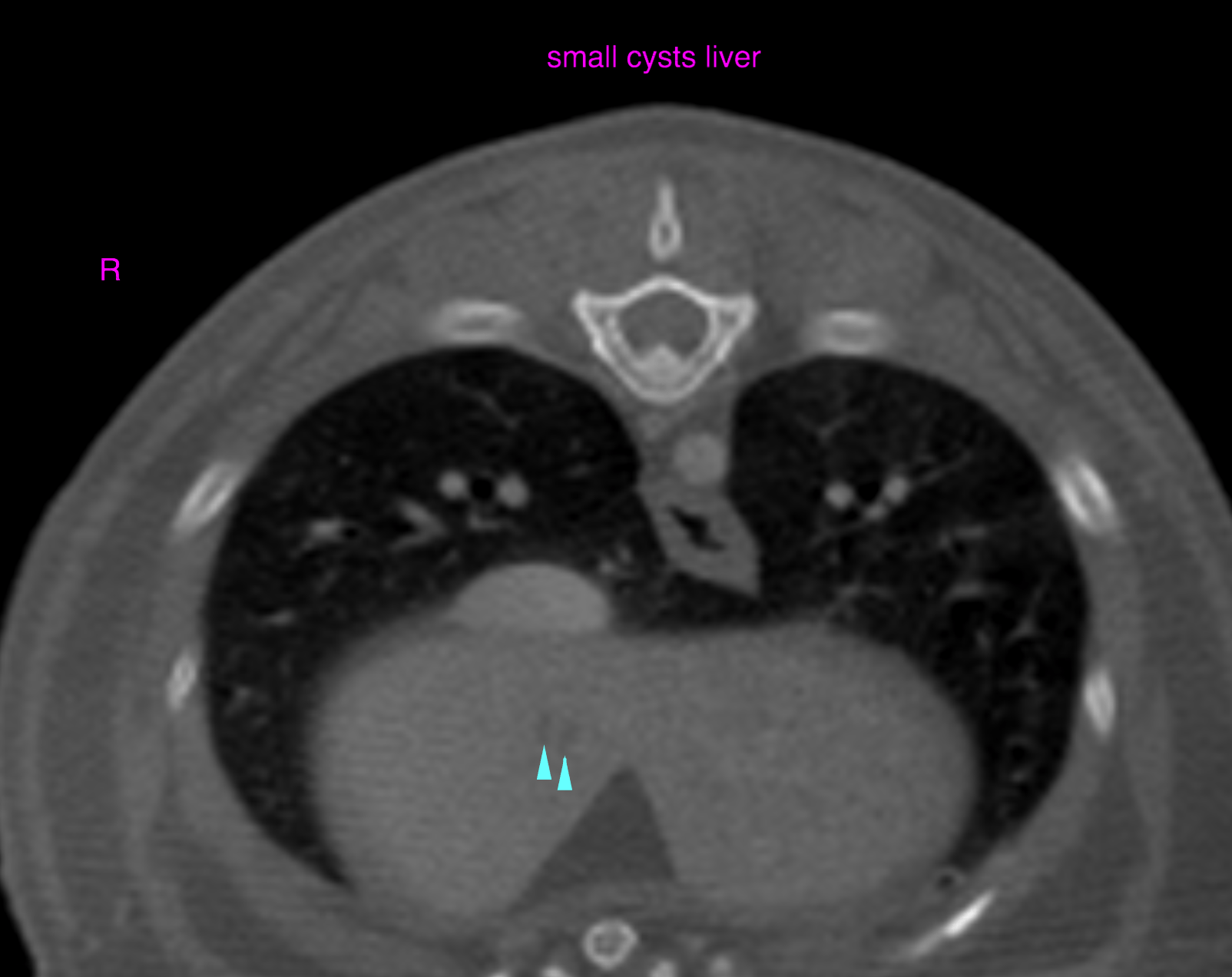
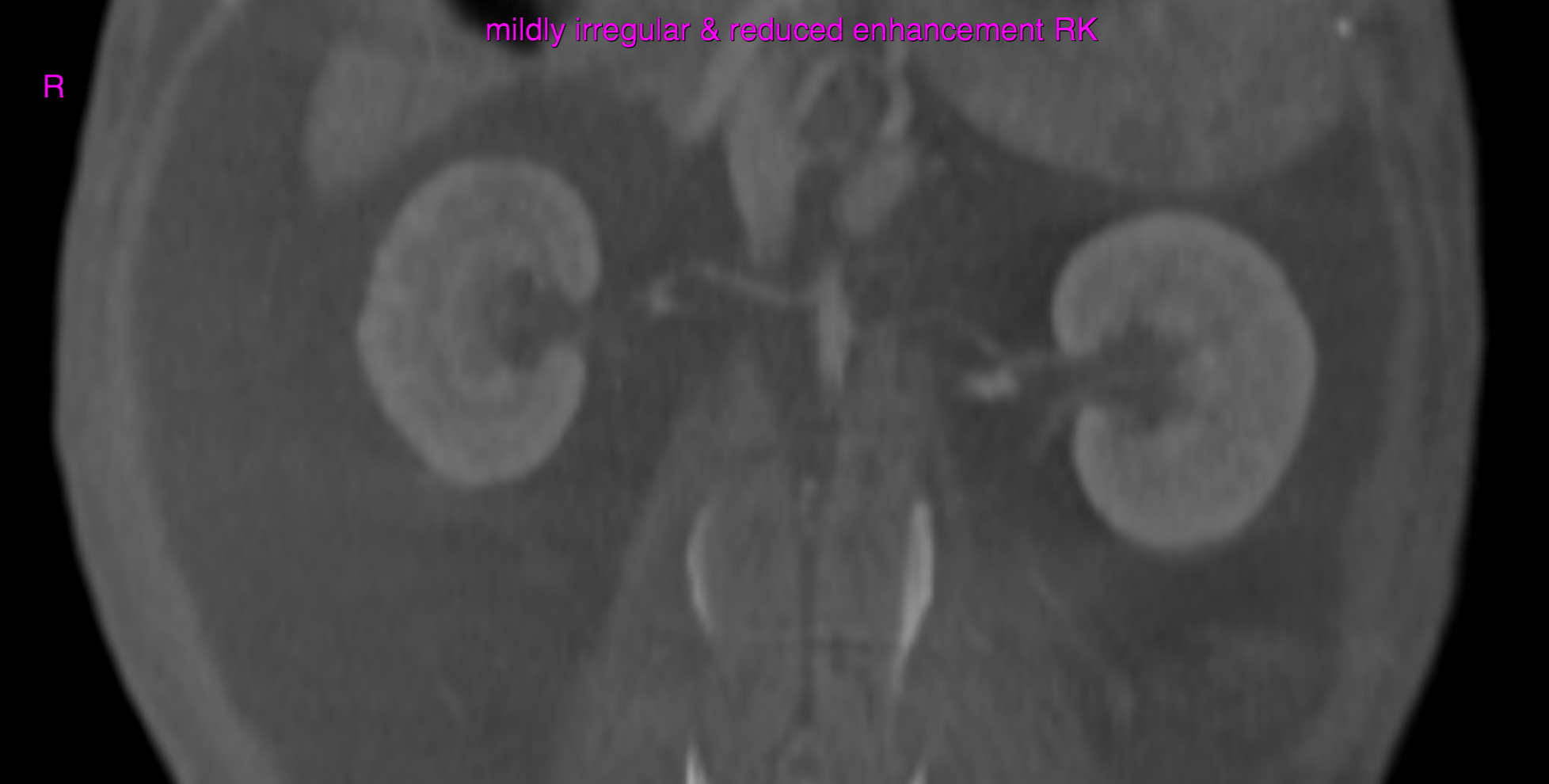
Both kidneys present a medullary rim sign. The right kidney is mildly irregular in shape and reveals decreased and slightly non-uniform contrast enhancement in the nephrogram phase. Several small cysts of < 5 mm with random distribution are noted within the liver parenchyma.
The spleen presents marked generalized enlargement with a patchy enhancement pattern which is likely to be due to the anesthesia related hypotension and differential enhancement of the red and white pulpa in the early phase of splenic perfusion.